Review
. 2015 Jul-Aug;51(4):257-67.
doi: 10.3109/10409238.2016.1172553. Epub 2016 Apr 20.

Ion channel-transporter interactions

Affiliations
Review

Ion channel-transporter interactions

Daniel L Neverisky et al. Crit Rev Biochem Mol Biol. 2015 Jul-Aug.

Abstract

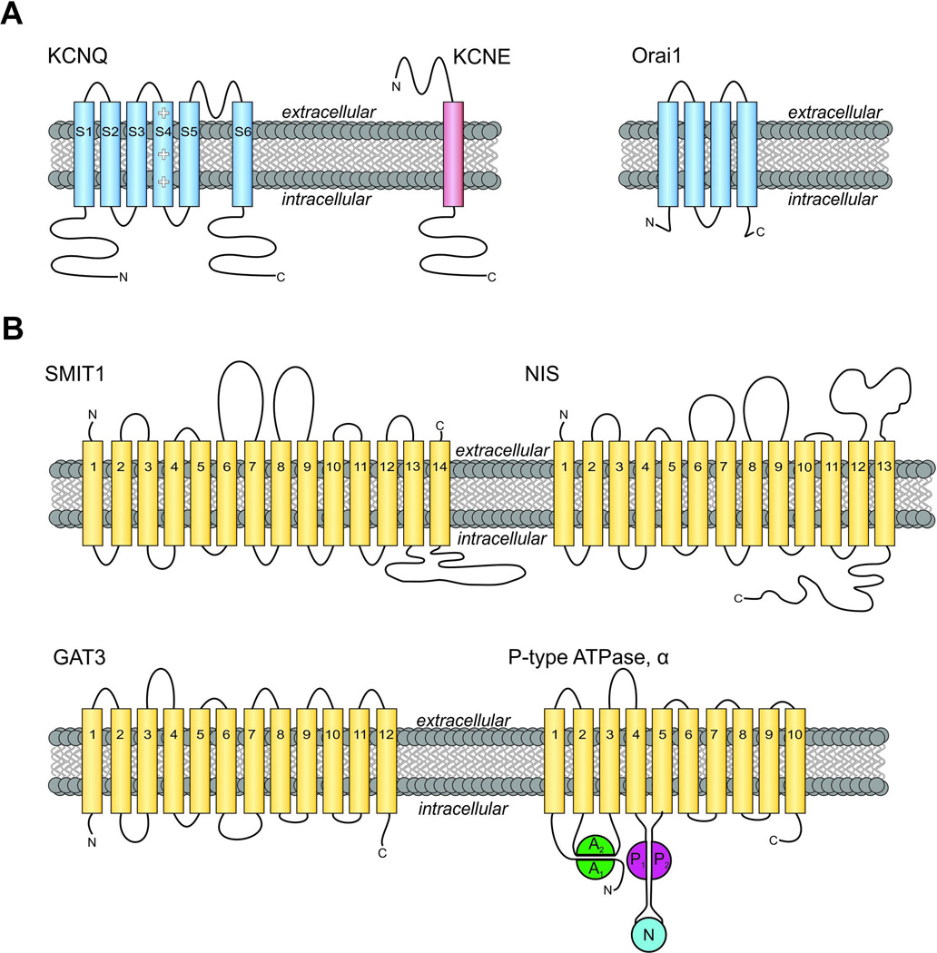
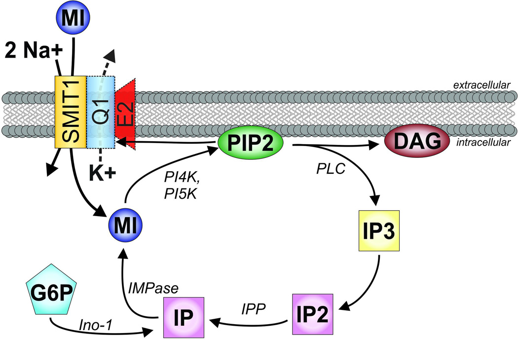
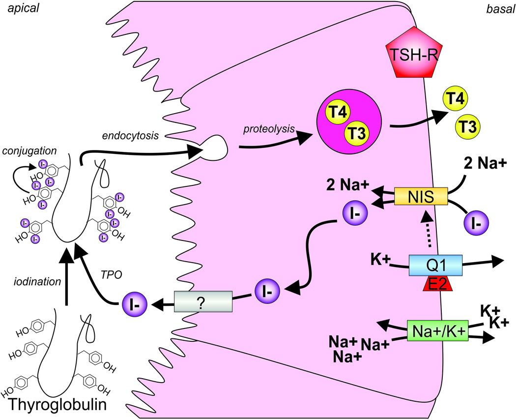
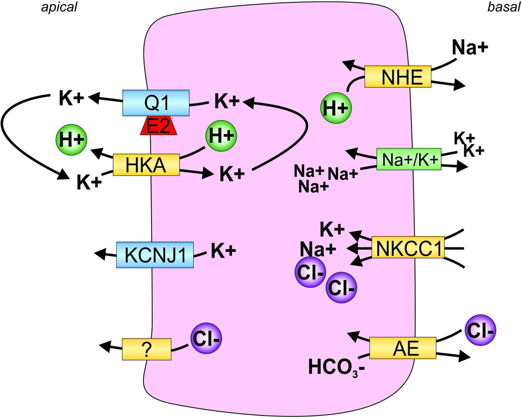
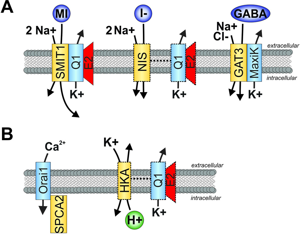
All living cells require membrane proteins that act as conduits for the regulated transport of ions, solutes and other small molecules across the cell membrane. Ion channels provide a pore that permits often rapid, highly selective and tightly regulated movement of ions down their electrochemical gradient. In contrast, active transporters can move moieties up their electrochemical gradient. The secondary active transporters (such as SLC superfamily solute transporters) achieve this by coupling uphill movement of the substrate to downhill movement of another ion, such as sodium. The primary active transporters (including H(+)/K(+)-ATPases and Na(+)/K(+)-ATPases) utilize ATP hydrolysis as an energy source to power uphill transport. It is well known that proteins in each of these classes work in concert with members of the other classes to ensure, for example, ion homeostasis, ion secretion and restoration of ion balance following action potentials. More recently, evidence is emerging of direct physical interaction between true ion channels, and some primary or secondary active transporters. Here, we review the first known members of this new class of macromolecular complexes that we term "chansporters", explore their biological roles and discuss the pathophysiological consequences of their disruption. We compare functional and/or physical interactions between the ubiquitous KCNQ1 potassium channel and various active transporters, and examine other newly discovered chansporter complexes that suggest we may be seeing the tip of the iceberg in a newly emerging signaling modality.

Keywords: ATPase; Active transport; KCNQ1; NIS; SMIT1; voltage-gated potassium channel.


Figures

Figure 1
Figure 1. Membrane topology of channel subunits and transporters discussed in this review
A. Left, transmembrane topology of KCNQ voltage-gated potassium (Kv) channel α subunits and KCNE β subunits. Kv pore-forming loop pictured between S5-S6. Right, topology of Orai1. Intracellular N- and C-terminals labeled accordingly. B. Upper, transmembrane topology of two SLC5A transporters. (Left) Sodium/myo-inositol co-transporter SMIT1, with extracellular N and C-terminals labeled accordingly. SMIT1 features a large intracellular loop between segments 13 and 14, as well as two large extracellular loops between segments 6–7 and 8–9. (Right) The sodium/iodide symporter NIS. Lower, transmembrane topology of GAT3 (SLC6A11) and a P-type ATPase α subunit such as the gastric H+/K+-ATPase. Extracellular N-terminus and intracellular C-terminus are labeled. Note that topology maps are shown for clarity and simplicity. High-resolution crystal structures have been solved for Orai (Hou et al. 2012), Kv1.2 (a mammalian relative of KCNQ channels) (Long et al. 2005), and vSGLT (a prokaryotic sodium galactose transporter related to mammalian SLC5A transporters) (Faham et al. 2008). Actual structures may deviate from predicted topologies with respect to, e.g., broken helices and in some cases actual transmembrane disposition. A color version of the figure is available online.
Figure 2
Figure 2. The myo-inositol pathway in the context of SMIT1 and KCNQ1-KCNE2
SMIT1, orange, transports sodium (Na+) and myo-inositol (MI) with a stoichiometry of 2:1. Phosphatidylinositol 4-kinase (PI4K), followed by phosphatidylinositol 5-kinase (PI5K), add phosphates to MI to form phosphatidylinositol 4,5-bisphosphate (PIP2). When activated, commonly by Gq-coupled receptors, phospholipase C (PLC) cleaves PIP2 into constituent components diacylglycerol (DAG) and inositol(1,4,5)-trisphosphate (IP3). IP3 is then recycled by dephosporylation to inositol(1,4)-bisphosphate (IP2) and inositol monophosphate (IP) by Inositol-1,4 bisphosphate 1-phosphatase (IPP), and back to (myo)inositol by inositol- 1(or 4)-monophosphatase (IMPase). IP can also be synthesized from glucose 6-phosphate (G6P) by inositol synthase (Ino-1). KCNQ1 (blue) with and without KCNE2 (red) can form complexes with SMIT1, although this interaction is not required for basic SMIT1 function. KCNQ1 gating is heavily regulated by PIP2. A color version of the figure is available online.
Figure 3
Figure 3. Channel-transporter crosstalk in the thyroid
The sodium-iodide symporter (NIS, orange) transports Na+ and I (purple) into the thyrocyte. This process is facilitated, via an incompletely resolved mechanism, by potassium ion efflux through KCNQ1-KCNE2 (Q1, blue; E2, red) channels and also involves active transport by the Na+/K+-ATPase (Na+/K+, green). I is then passed through to the colloid by another protein (?) whose identity is still under debate. I undergoes oxidation by thyroid peroxidase (TPO) and is incorporated into the thyroglobulin (Tg) molecule to produce monoiodotyrosine (MIT) and diiodotyrosine (DIT) by organification. The organified Tg is then resorbed by the follicular cells where, when the thyroid stimulating hormone receptor is activated (TSH-R, red), the iodinated Tgs are cleaved to form either thyroxine (T4), generated from a pair of DIT, or triiodothyronine (T3), produced from one MIT and one DIT molecule. A color version of the figure is available online.
Figure 4
Figure 4. Channel-transporter crosstalk in gastric parietal cells facilitates gastric acid secretion
Sodium-hydrogen exchanger (NHE) draws Na+ into the cell and expels H+. The sodium-potassium ATPase (Na+/K+) releases Na+ and draws in K+. Na+/K+/Cl cotransporter 1 (NKCC1) draws Na+, K+, and Cl into the cell. The anion exchanger (AE) exchanges Cl and HCO3. H+/K+-ATPase (HKA) expels H+ into the stomach lumen accompanied by an unknown Cl channel or transporter to participate in gastric acid formation. KCNQ1-KCNE2 (Q1, E2) and KCNJ channels expel potassium from the cell in order to “reset” the high intracellular K+ accumulated by HKA activity. A color version of the figure is available online.
Figure 5
Figure 5. Summary of channel-transporter complex formation and functional crosstalk discussed herein
A. K+ channel interactions with members of the SLC superfamily. Left, SMIT1 and KCNQ1-KCNE2 form reciprocally regulating, physically interacting complexes in the brain. Center, NIS and KCNQ1-KCNE2 exhibit functional crosstalk in the thyroid, although physical interaction remains to be determined (indicated by dotted lines). Right, GAT3 and MaxiK form physically interacting chansporter complexes. B. Left, Orai1 and SPCA2 form physically interacting chansporter complexes which enable a novel mode of calcium channel activation, potentially important in tumorigenesis. Right, HKA and KCNQ1-KCNE2 exhibit functional crosstalk in the stomach to enable gastric acid secretion. Physical interaction remains to be determined (indicated by dotted lines). A color version of the figure is available online.

References

    1. Abbott GW. Biology of the KCNQ1 Potassium Channel. [Accessed June 4, 2014];New Journal of Science. 2014 2014:1–26. Available at: http://www.hindawi.com/journals/njos/2014/237431/
    1. Abbott GW, et al. KCNQ1, KCNE2, and Na+-coupled solute transporters form reciprocally regulating complexes that affect neuronal excitability. [Accessed May 31, 2014];Science signaling. 2014 7(315):ra22. Available at: http://www.ncbi.nlm.nih.gov/pubmed/24595108. - PMC - PubMed
    1. Abbott GW, et al. MiRP2 Forms Potassium Channels in Skeletal Muscle with Kv3 . 4 and Is Associated with Periodic Paralysis. Channels. 2001;104:217–231. - PubMed
    1. Allison JH, Stewart MA. Reduced Brain Inositol in Lithium-treated Rats. Nature New Biology. 1971;233(43):267–268. Available at: http://www.nature.com/doifinder/10.1038/newbio233267a0. - DOI - PubMed
    1. Angelo K, et al. KCNE5 induces time- and voltage-dependent modulation of the KCNQ1 current. Biophysical journal. 2002;83(4):1997–2006. Available at: http://dx.doi.org/10.1016/S0006-3495(02)73961-1. - DOI - PMC - PubMed

Publication types