Multicenter Study
. 2016 Jun 23;127(25):3281-90.
doi: 10.1182/blood-2015-12-684498. Epub 2016 Apr 20.

The minimum required level of donor chimerism in hereditary hemophagocytic lymphohistiocytosis

Affiliations
Multicenter Study

The minimum required level of donor chimerism in hereditary hemophagocytic lymphohistiocytosis

Bernd Hartz et al. Blood. .

Abstract

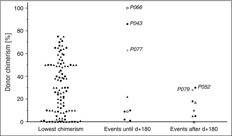
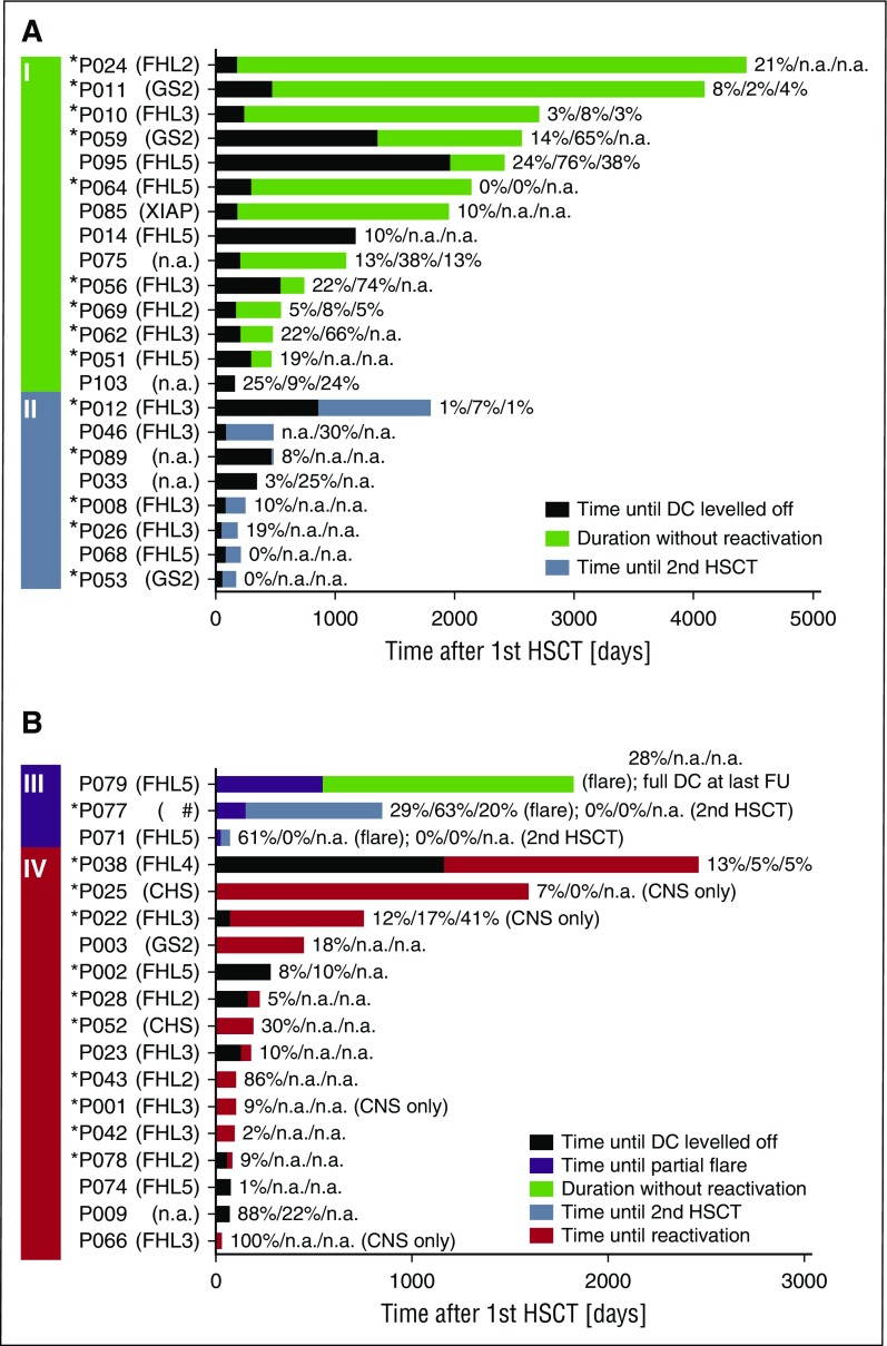
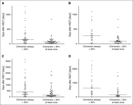
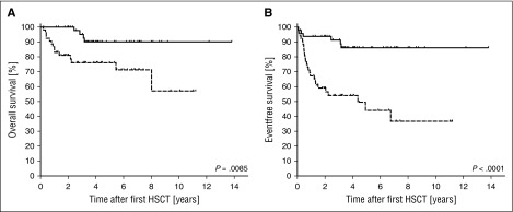
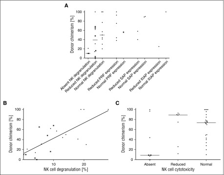
Reduced-intensity conditioning has improved survival after hematopoietic stem cell transplantation (HSCT) for hemophagocytic lymphohistiocytosis (HLH) at the cost of more frequent mixed chimerism. The minimum level of donor chimerism (DC) required to prevent HLH reactivation in humans remains to be determined. In a multicenter retrospective study, 103 patients transplanted for hereditary HLH (2000-2013) and DC permanently or transiently <75% (overall, CD3(+), CD56(+)) were analyzed regarding DC, specific immunologic function, occurrence of systemic reactivations (≥5/8 HLH criteria), partial systemic flares (<5 criteria and HLH-directed treatment), isolated central nervous system reactivations, and management. Recurrence was reported in 18 patients (systemic reactivation n = 11, partial flare n = 3, isolated central nervous system reactivation n = 4). Ten events occurred during profound immune suppression before day 180 (median DC, 10%; range, 1-100%; CD3(+) if available, otherwise overall DC), which renders a differentiation between secondary post-HSCT HLH and HLH related to the genetic defect difficult. Eight events occurred between 0.5 and 6.7 years post-HSCT (median DC, 13%; range, 0-30%). In 5 patients, overall and lineage-specific DC were ≤10% for >6 months (median, 5.1; range, 1.1-10 years) without reactivation. A second HSCT was performed in 18 patients (median, DC 4%; range, 0-19%). Death from reactivation occurred in 4 patients (22% of recurrences). Six patients died of transplant complications following a second HSCT (33% of second HSCT). We conclude that a DC >20%-30% is protective against late reactivation. Lower levels do not, however, inescapably result in recurrences. The decision for or against second HSCT must be based on a thorough risk assessment.


Figures

Figure 1
Figure 1
DC in systemic and CNS reactivations and partial flares. The level of DC is depicted, if available, as CD3+ DC, or otherwise as overall DC. Left: Lowest DC level of each patient (CD3+, ▲; overall, ●). Middle and right: Patients with systemic (CD3+, ▲; overall, ●), isolated CNS reactivations (CD3+, △; overall, ○), and partial flares (CD3+, ▲; overall, ●) at the time of the event. Events in the middle column occurred during profound post-HSCT immune suppression before day 180; events in the right column occurred after that day. Refer to the “Results” section for case descriptions of events that occurred at a DC >25%. Patient identifications are indicated in the figure.
Figure 2
Figure 2
Timeline after first HSCT. Four groups of patients are included in this figure. (A) Patients who never experienced a reactivation or partial flare, did not have a second HSCT, and had a DC of ≤30% at last follow-up (I). Note that long-term event-free survival was possible in this group despite low DC levels. Patients who did not experience a reactivation or partial flare and received a second HSCT because of low DC (II). (B) Patients with a partial flare (III). Patients with systemic or isolated CNS reactivation (IV). The black part of the bar displays the time from first HSCT until the date when DC leveled off (ie, DC did not vary for >±10% anymore) and the following time until (I) last follow-up (green), (II) second HSCT (blue), (III) partial flare (purple), last follow-up (green), or second HSCT (blue), or (IV) reactivation (red). In some patients, the time when DC leveled off was after an event and is thus not shown. The DC (overall/CD3+/CD56+) at last follow-up (I), at second HSCT (II), at partial flare and last follow-up or second HSCT (III), and reactivation (IV) is provided. In P064, the absent DC for 5 years is shown, even though it became detectable again at last measurement (see the “Results” section for case description). *Patients with early-onset HLH during infancy (≤12 months of age), indicating a severe genetic defect. #A double heterozygous defect was found in this patient, 1 each for FHL3 and 5. CD3, T cell; CD56, NK cell; NA, not available.
Figure 3
Figure 3
Dynamics of loss of donor chimerism. The time interval is shown from first HSCT until overall (A) and CD3+ (B) DC fell below 75% and until overall (C) and CD3+ (D) DC reached the nadir. The intervals were significantly shorter in patients in whom the lowest DC was ≤30% compared with patients who always had a DC >30%. (A) P < .0001; (B) P = .0002; (C) P = .0018; (C) P = .0002.
Figure 4
Figure 4
Kaplan-Meier estimated overall and event-free survival after first HSCT. Overall survival (A) and event-free survival (B) were significantly better in the group of 50 patients in which the DC was always >30% (solid line) than in the group of 53 patients in which the lowest DC level was ≤30% (dashed line). The respective hazard ratios were 3.5 and 4.6. An event was defined as systemic or isolated CNS reactivation, partial flare, death, or second HSCT.
Figure 5
Figure 5
Protein expression and NK cytotoxicity after HSCT. Functional analyses were correlated with the level of DC. Because the depicted assays had been performed with NK cells, CD56+ DC (□) is shown if available, otherwise the overall DC (●) is shown. (A) The results of the NK CD107a degranulation assay without interleukin-2 stimulation (FHL3-5, GS2, and CHS), expression of perforin (FHL2), SAP (XLP1), and XIAP (XLP2) were grouped into absent, reduced, and normal, according to local laboratory standards. (B) The percentage of degranulating (CD107a+) cells and the DC show a significant positive correlation (Spearman P = .005, ρ = .71). (C) NK cytotoxicity results were grouped into absent, reduced, and normal according to local laboratory standards. There was substantial overlap of DC level between the groups (P = .11). PRF, perforin.

Similar articles

Cited by

References

    1. Henter JI, Horne A, Aricó M, et al. HLH-2004: diagnostic and therapeutic guidelines for hemophagocytic lymphohistiocytosis. Pediatr Blood Cancer. 2007;48(2):124–131. - PubMed
    1. Janka GE, Lehmberg K. Hemophagocytic syndromes—an update. Blood Rev. 2014;28(4):135–142. - PubMed
    1. Baker KS, Filipovich AH, Gross TG, et al. Unrelated donor hematopoietic cell transplantation for hemophagocytic lymphohistiocytosis. Bone Marrow Transplant. 2008;42(3):175–180. - PubMed
    1. Horne A, Janka G, Maarten Egeler R, et al. Histiocyte Society. Haematopoietic stem cell transplantation in haemophagocytic lymphohistiocytosis. Br J Haematol. 2005;129(5):622–630. - PubMed
    1. Ouachée-Chardin M, Elie C, de Saint Basile G, et al. Hematopoietic stem cell transplantation in hemophagocytic lymphohistiocytosis: a single-center report of 48 patients. Pediatrics. 2006;117(4):e743–e750. - PubMed

Publication types

MeSH terms