Review
. 2017 Apr;32(4):547-555.
doi: 10.1007/s00467-016-3372-y. Epub 2016 Apr 21.

Xenopus: leaping forward in kidney organogenesis

Affiliations
Review

Xenopus: leaping forward in kidney organogenesis

Vanja Krneta-Stankic et al. Pediatr Nephrol. 2017 Apr.

Abstract

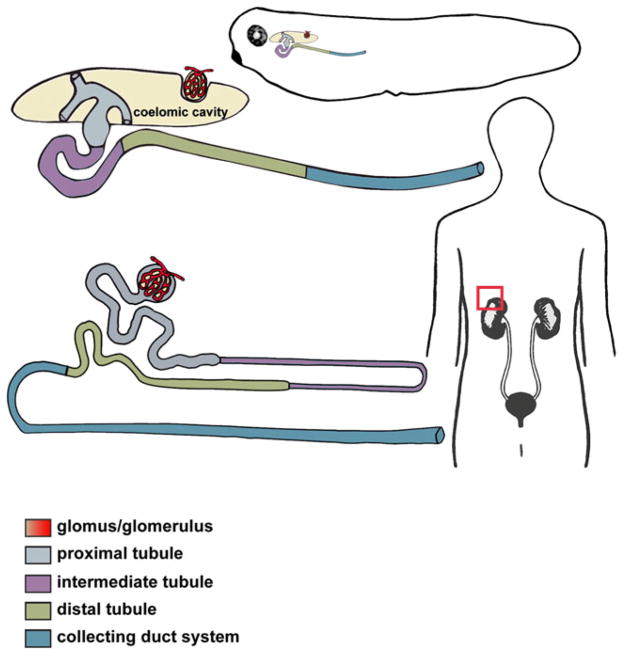
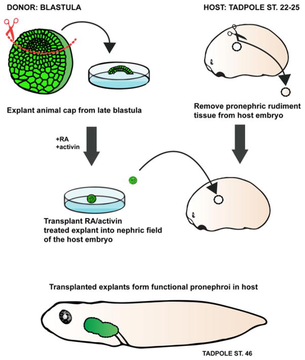
While kidney donations stagnate, the number of people in need of kidney transplants continues to grow. Although transplanting culture-grown organs is years away, pursuing the engineering of the kidney de novo is a valid means of closing the gap between the supply and demand of kidneys for transplantation. The structural organization of a mouse kidney is similar to that of humans. Therefore, mice have traditionally served as the primary model system for the study of kidney development. The mouse is an ideal model organism for understanding the complexity of the human kidney. Nonetheless, the elaborate structure of the mammalian kidney makes the discovery of new therapies based on de novo engineered kidneys more challenging. In contrast to mammals, amphibians have a kidney that is anatomically less complex and develops faster. Given that analogous genetic networks regulate the development of mammalian and amphibian nephric organs, using embryonic kidneys of Xenopus laevis (African clawed frog) to analyze inductive cell signaling events and morphogenesis has many advantages. Pioneering work that led to the ability to generate kidney organoids from embryonic cells was carried out in Xenopus. In this review, we discuss how Xenopus can be utilized to compliment the work performed in mammalian systems to understand kidney development.

Keywords: Development; Induction; Kidney; Nephron; Organoid; Pronephros; Xenopus.


Conflict of interest statement

Conflict of interest statement: The authors declare that they have no conflicts of interest

Figures

Fig. 1
Fig. 1
Xenopus pronephric and human metanephric nephrons share a conserved segmentation pattern. A schematic representation of an enlarged Xenopus pronephric nephron (top) and mammalian metanephric nephron (bottom), with the distinct tubular compartments labeled. The glomus/glomerulus filters the fluid from the blood plasma through capillary walls into the proximal tubule. The drawing depicts a slight difference in organization of the glomar/glomerular anatomy between the amphibian pronephric and mammalian metanephric nephrons. In mammalian metanephric nephrons, the glomerulus is integrated into the Bowman’s capsule near the proximal tubule, while the Xenopus glomus projects into a body cavity (coelomic cavity). Three branches of pronephric tubule (nephrostomes) use ciliary action to force the glomar filtrate down the pronephric tubule. Together, the tubule segments (proximal, intermediate and distal) and the collecting duct system (connecting tubule and duct) facilitate the process of filtrate reabsorption and transport of wastes products for secretion
Fig. 2
Fig. 2
Xenopus in vitro-induced kidney organoids. Isolated animal cap (presumptive ectodermal tissue) explants (green) treated with activin and retinoic acid (RA) differentiate to form pronephric organoids in culture. The animal cap tissue treated with activin and RA can be transplanted into a host embryo which lacks kidney primordia. The in vitro-induced pronephric tissue is capable of functioning in vivo and compensates for the loss of the host kidneys by suppressing edema formation
Fig. 3
Fig. 3
In vivo imaging of the frog pronephros. Xenopus fate-map of early development permits microinjections of blastomeres fate-mapped to kidney cell progenitors and it can be utilized for targeted delivery of fluorophores to developing pronephros. Microinjected embryos can be immobilized in low melting-point agarose and subjected to time-lapse imaging. This sample preparation allows for in vivo analysis of cell behaviors and molecular dynamics underlying pronephros formation

References

    1. Moody SA. Fates of the blastomeres of the 16-cell stage Xenopus embryo. Dev Biol. 1987;119:560–578. - PubMed
    1. Moody SA. Fates of the blastomeres of the 32-cell stage Xenopus embryo. Dev Biol. 1987;122:300–319. - PubMed
    1. Bauer DV, Huang S, Moody SA. The cleavage stage origin of Spemann’s organizer: analysis of the movements of blastomere clones before and during gastrulation in Xenopus. Development. 1994;120:1179–1189. - PubMed
    1. DeLay BD, Krneta-Stankic V, Miller RK. Technique to target microinjection to the developing Xenopus kidney. J Vis Exp. 2016:e53799. doi: 10.3791/53799. - DOI - PMC - PubMed
    1. Saxén L. Organogenesis of the kidney. Cambridge University Press; Cambridge: 1987.

Publication types

LinkOut - more resources