. 2016 May;28(5):1200-14.
doi: 10.1105/tpc.16.00007. Epub 2016 Apr 20.

UBIQUITIN-SPECIFIC PROTEASE14 Interacts with ULTRAVIOLET-B INSENSITIVE4 to Regulate Endoreduplication and Cell and Organ Growth in Arabidopsis

Affiliations

UBIQUITIN-SPECIFIC PROTEASE14 Interacts with ULTRAVIOLET-B INSENSITIVE4 to Regulate Endoreduplication and Cell and Organ Growth in Arabidopsis

Yingxiu Xu et al. Plant Cell. 2016 May.

Abstract

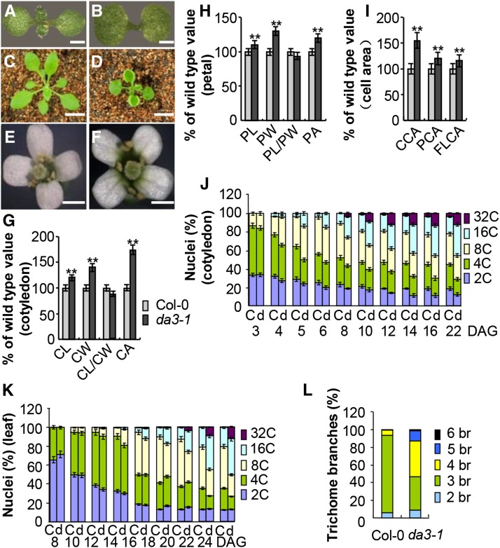
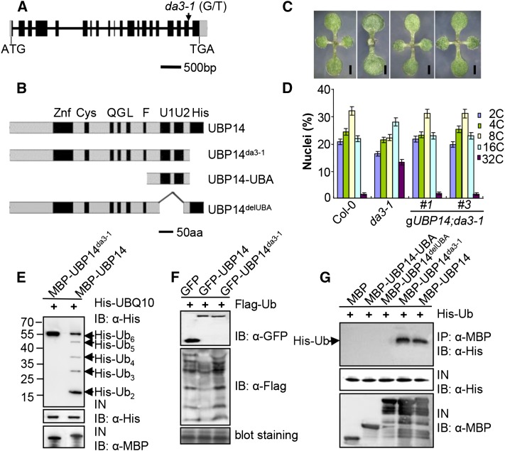
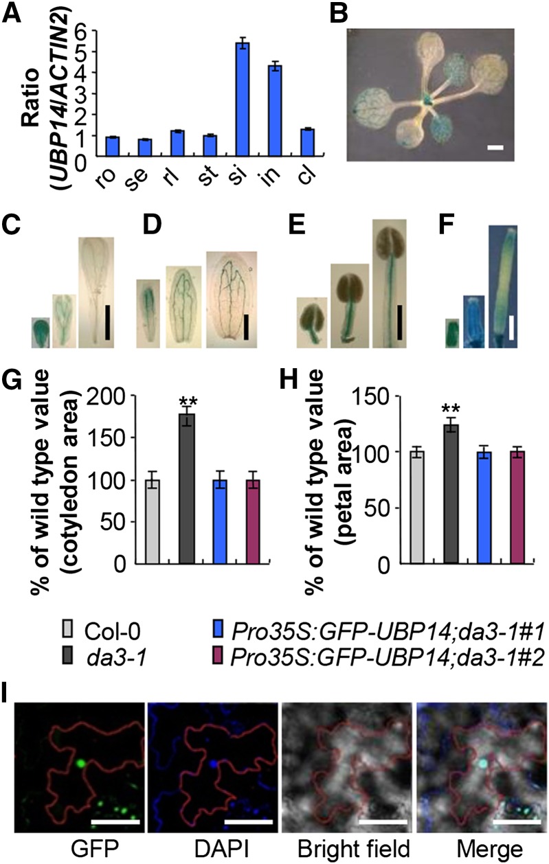
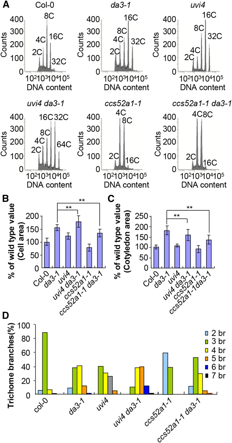
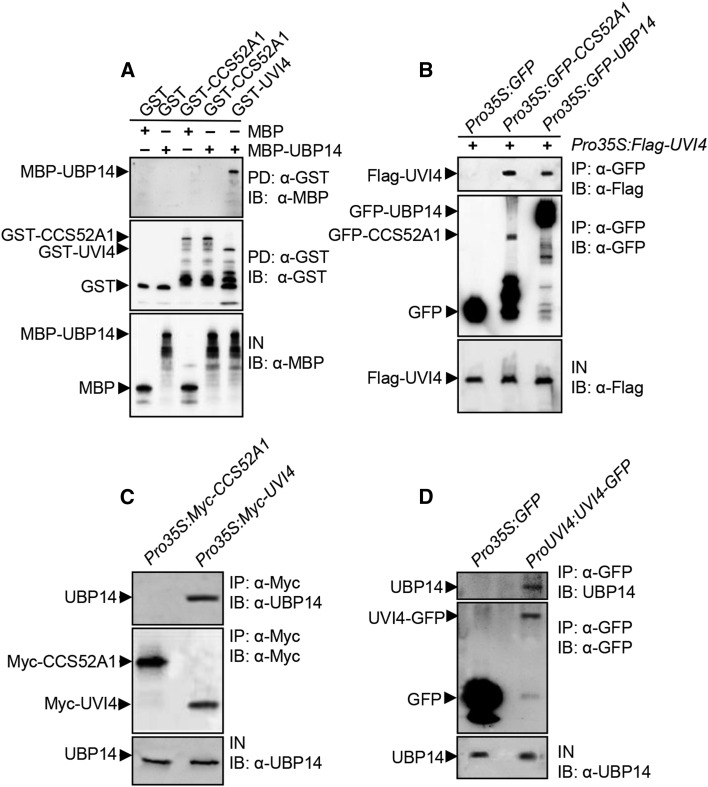
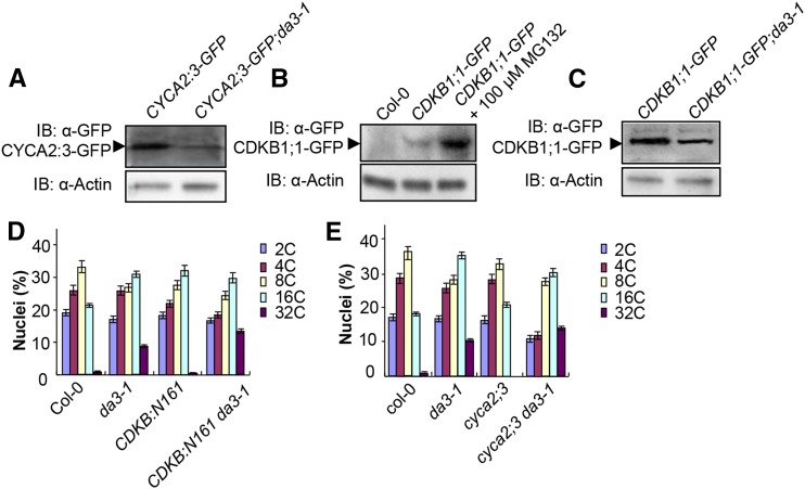
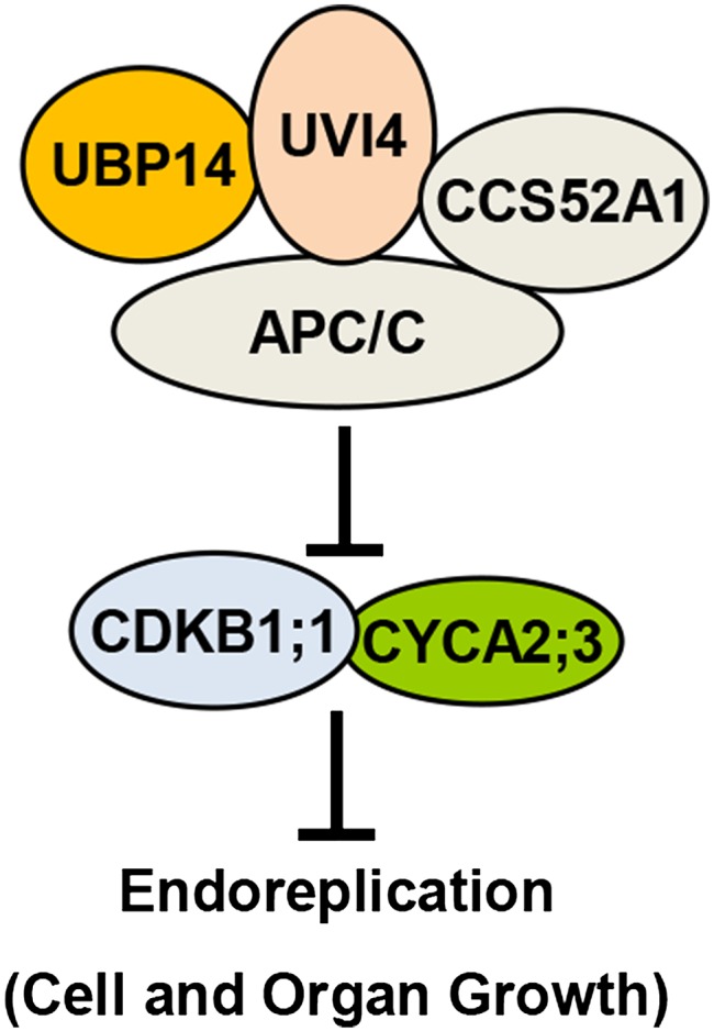
Organ growth is determined by a coordinated combination of cell proliferation and cell growth and differentiation. Endoreduplication is often coupled with cell growth and differentiation, but the genetic and molecular mechanisms that link endoreduplication with cell and organ growth are largely unknown. Here, we describe UBIQUITIN-SPECIFIC PROTEASE14 (UBP14), encoded by the DA3 gene, which functions as a negative regulator of endoreduplication. The Arabidopsis thaliana da3-1 mutant shows large cotyledons, leaves, and flowers with higher ploidy levels. UBP14 acts along with UV-B-INSENSITIVE4 (UVI4), an inhibitor of the anaphase-promoting complex/cyclosome (APC/C) ubiquitin ligase, to repress endoreduplication. Also, UBP14 functions antagonistically with CELL CYCLE SWITCH52 A1 (CCS52A1), an activator of APC/C, to regulate endoreduplication. UBP14 physically associates with UVI4 both in vitro and in vivo but does not directly interact with CCS52A1. Further results reveal that UBP14 influences the stability of cyclin A2;3 (CYCA2;3) and cyclin-dependent kinase B1;1 (CDKB1;1), two downstream components of the APC/C Thus, our findings show how endoreduplication is linked with cell and organ growth by revealing important genetic and molecular functions for the ubiquitin-specific protease UBP14 and for the key cell cycle regulators UVI4, CCS52A1, CYCA2;3, and CDKB1;1.


Figures

Figure 1.
Figure 1.
da3-1 Influences Endoreduplication and Cell and Organ Growth. (A) and (B) Eight-day-old seedlings of Col-0 (A) and da3-1(B). (C) and (D) Twenty-four-day-old seedlings of Col-0 (C) and da3-1(D). (E) and (F) Flowers of Col-0 (E) and da3-1(F). (G) Cotyledon length (CL), cotyledon width (CW), cotyledon length/cotyledon width, and cotyledon area (CA) of 10-d-old Col-0 and da3-1 seedlings (n = 60). (H) Petal length (PL), petal width (PW), petal length/petal width, and petal area (PA) of Col-0 and da3-1 flowers (stage 14) (n = 50). (I) The average area of cells in cotyledons (CCA) of 10-d-old Col-0 and da3-1 seedlings, petals (PCA) (stage 14), and the first pair of leaves (FLCA) of 21-d-old Col-0 and da3-1 seedlings . Thirty-five cotyledons (n = 35), thirty-five petals (n = 35), and thirty leaves (n = 30) were used to measure cell area. (J) Nuclear DNA ploidy distribution of cells in Col-0 (C) and da3-1(D) cotyledons over a period of 22 DAG (n = 3). Cotyledons were harvested at the indicated times and ploidy was determined by flow cytometry. (K) Nuclear DNA ploidy distribution of cells in the first pair of leaves of Col-0 (C) and da3-1(D) seedlings over a period of 24 DAG (n = 3). The first pair of leaves was harvested at the indicated times and ploidy was determined by flow cytometry. (L)Trichome branch (br) distribution of Col-0 and da3-1 in the first pair of leaves of seedlings at 15 DAG (n = 200). Values in (G) to (K) are given as means ± sd relative to the values for the respective wild-type seedlings, set at 100%. **P < 0.01 compared with the wild type (Student’s t test). Bars =1 mm in (A) and (B), 2 cm in (C) and (D), and 1 mm in (E) and (F).
Figure 2.
Figure 2.
DA3 Encodes the Ubiquitin-Specific Protease UBP14. (A) DA3/UBP14 structure. The start codon (ATG) and the stop codon (TGA) for this gene are indicated. Black boxes indicate the coding sequence, gray boxes indicate the 5′ and 3′ untranslated regions, and lines between boxes indicate introns. The mutation site of da3-1 is shown by an arrow. (B) DA3/UBP14 structure. This protein contains the Znf, Cys, His, Q, G, and L motifs and two ubiquitin-associated domains (U1 and U2). Schematic diagrams of UBP14, UBP14da3-1, and its deletion mutants are shown. aa, amino acids. (C) Ten-day-old seedlings of Col-0, da3-1, gUBP14;da3-1#1, and gUBP14;da3-1#3 (from left to right). gUBP14;da3-1 represents da3-1 transformed with the genomic sequence of the UBP14 gene. Bars = 1 mm. (D) Nuclear DNA ploidy distribution of cells in Col-0, da3-1, gUBP14;da3-1#1, and gUBP14;da3-1#3 cotyledons (n = 3). gUBP14;da3-1 represents da3-1 transformed with the genomic sequence of UBP14. Each bar represents the mean ± sd of three replicates. (E) UBP14 has deubiquitination activity in vitro. MBP-UBP14 cleaved His-UBQ10 in vitro, but MBP-UBP14da3-1 did not. His-UBQ10 and cleaved His-UBQ10 proteins were detected by immunoblot analysis with an anti-His antibody. (F) UBP14 has deubiquitination activity in vivo. N. benthamiana leaves were transformed by injection of Agrobacterium GV3101 cells harboring Pro35S:Flag-UBQ14 with Pro35S:GFP, Pro35S:GFP-UBP14, or Pro35S:GFP-UBP14da3-1 plasmids, respectively. Total ubiquitinated proteins were detected by immunoblot analysis with an anti-Flag antibody. (G) UBA motifs are required for ubiquitin binding. His-ubiquitin was pulled down by MBP, MBP-UBP14-UBA, MBP-UBP14delUBA, MBP-UBP14da3-1, or MBP-UBP14 and then immobilized on amylose resin and analyzed by immunoblotting using an anti-His antibody.
Figure 3.
Figure 3.
Expression Pattern and Subcellular Localization of UBP14. (A) Quantitative real-time RT-PCR analysis of DA3 expression in the indicated Arabidopsis tissues. Total RNA was isolated from roots (ro), seedlings (se), rosette leaves (rl), stems (st), siliques (si), inflorescences (in), and cauline leaves (cl). Data shown are means ± sd of three biological replicates. (B) to (F) UBP14 expression activity was monitored by ProUBP14:GUS transgene expression. Twenty GUS-expressing lines were observed, and all showed a similar pattern, although they differed slightly in the intensity of the staining. Histochemical analysis of GUS activity in 12-d-old seedlings (B), the developing petals (C), the developing sepals (D), the developing stamens (E), and the developing carpels (F). (G) Cotyledon area of seedlings of Col-0, da3-1, Pro35S:GFP-UBP14;da3-1#1, and Pro35S:GFP-UBP14;da3-1#2 (n = 55). Pro35S:GFP-UBP14;da3-1 represents da3-1 transformed with Pro35S:GFP-UBP14. (H) Petal area of Col-0, da3-1, Pro35S:GFP-UBP14;da3-1#1, and Pro35S:GFP-UBP14;da3-1#2 (n = 65). Pro35S:GFP-UBP14;da3-1 is da3-1 transformed with Pro35S:GFP-UBP14. (I) Subcellular localization of GFP-UBP14 in epidermal cells of Pro35S:GFP-UBP14;da3-1 leaves. GFP fluorescence of GFP-UBP14, DAPI staining, and bright-field and merged images (from left to right) are shown. The red lines indicate the outline of an epidermal cell. Values in (G) and (H) are given as means ± sd relative to the respective wild-type values, set at 100%. **P < 0.01 compared with the wild type (Student’s t test). Bars = 1 mm in (B), 1 mm in (C) to (F), and 50 μm in (I).
Figure 4.
Figure 4.
Genetic Analyses of UBP14 with UVI4 and CCS52A1 in Regulation of Endoreduplication. (A) Flow cytometric analysis of cells in Col-0, da3-1, uvi4, uvi4 da3-1, ccs52a1-1, and ccs52a1-1 da3-1 cotyledons. (B) The average area of palisade cells in Col-0, da3-1, uvi4, uvi4 da3-1, ccs52a1-1, and ccs52a1-1 da3-1 cotyledons. Thirty cotyledons were used to measure cell area (n = 30). (C) The average area of cotyledons of Col-0, da3-1, uvi4, uvi4 da3-1, ccs52a1-1, and ccs52a1-1 da3-1 (n = 50). (D)Trichome branch (br) distribution in the first pair of leaves of Col-0, da3-1, uvi4, uvi4 da3-1, ccs52a1-1, and ccs52a1-1 da3-1 seedlings at 15 DAG (n = 200). Values in (B) and (C) are given as means ± sd relative to the respective wild-type values, set at 100%. **P < 0.01 compared with the wild type (Student’s t test).
Figure 5.
Figure 5.
UBP14 Physically Interacts with UVI4. (A) UBP14 physically interacts with UVI4 in vitro. MBP-UBP14 was pulled down (PD) by GST, GST-CCS52A1, or GST-UVI4 immobilized on GST beads and analyzed by immunoblotting (IB) using an anti-MBP antibody. IN represents input. (B) UBP14 interacts with UVI4 in vivo. N. benthamiana leaves were transformed by injection of Agrobacterium GV3101 cells harboring Pro35S:Flag-UVI4 and Pro35S:GFP, Pro35S:GFP-CCS52A1, or Pro35S:GFP-UBP14 plasmids. Total proteins were immunoprecipitated (IP) with GFP-Trap-A, and the immunoblot was probed with anti-GFP and anti-Flag antibodies, respectively. (C) UBP14 associates with UVI4 in Arabidopsis. Total proteins from Pro35S:Myc-CCS52A1 or Pro35S:Myc-UVI4 seedlings were immunoprecipitated with anti-Myc-Tag mouse mAb conjugated agarose beads, and the immunoblot was probed with anti-UBP14 and anti-Myc antibodies, respectively. (D) UBP14 associates with UVI4 in Arabidopsis. Total proteins from Pro35S:GFP or ProUVI4:UVI4-GFP seedlings were immunoprecipitated with GFP-Trap-A, and the immunoblot was probed with anti-UBP14 and anti-GFP antibodies, respectively.
Figure 6.
Figure 6.
UBP14 Modulates the Stability of CYCA2;3 and CDKB1;1. (A) The da3-1 mutation decreases the level of CYCA2;3-GFP. Total proteins from CYCA2;3-GFP and CYCA2;3-GFP;da3-1 seedlings induced with 50 μM β-estradiol for 16 h were subjected to immunoblot (IB) assays using anti-GFP and anti-actin antibodies, with actin used as the loading control. (B) The proteasome inhibitor MG132 stabilizes CDKB1;1. Ten-day-old CDKB1;1-GFP seedlings were treated with or without 100 μM MG132. Col-0 seedlings were used as a negative control. Total protein extracts were subjected to immunoblot assays using anti-GFP and anti-actin antibodies. (C) The da3-1 mutation decreases the level of CDKB1;1-GFP. Total proteins from CDKB1;1-GFP and CDKB1;1-GFP;da3-1 seedlings were subjected to immunoblot assays using anti-GFP and anti-actin antibodies. (D) Nuclear DNA ploidy distribution of cells in Col-0, da3-1, CDKB1;1N161, and CDKB1;1N161 da3-1 cotyledons at 10 DAG as analyzed by flow cytometry (n = 3). (E) Nuclear DNA ploidy distribution of cells in Col-0, da3-1, cyca2;3, and cyca2;3 da3-1 cotyledons at 10 DAG as analyzed by flow cytometry (n = 3). Values in (D) and (E) are given as means ± sd of three replicates.
Figure 7.
Figure 7.
A Model for UBP14 Functions in Endoreduplication and Cell and Organ Growth. UBP14 physically interacts with UVI4, an inhibitor of the APC/C ubiquitin ligase. UBP14 and UVI4 may work together to repress the activity of APC/C. It is also possible that UBP14 may function redundantly or in parallel with UVI4 to inhibit the activity of APC/C. UBP14 and UVI4 function antagonistically with CCS52A1 to regulate the activity of APC/C. APC/C has been shown to regulate the stability of CYCA2;3 and CDKB1;1. UBP14 influences the abundance of CYCA2;3 and CDKB1;1, which repress endoreduplication.

Similar articles

Cited by

References

    1. Baloban M., Vanstraelen M., Tarayre S., Reuzeau C., Cultrone A., Mergaert P., Kondorosi E. (2013). Complementary and dose-dependent action of AtCCS52A isoforms in endoreduplication and plant size control. New Phytol. 198: 1049–1059. - PubMed
    1. Boudolf V., Vlieghe K., Beemster G.T., Magyar Z., Torres Acosta J.A., Maes S., Van Der Schueren E., Inzé D., De Veylder L. (2004). The plant-specific cyclin-dependent kinase CDKB1;1 and transcription factor E2Fa-DPa control the balance of mitotically dividing and endoreduplicating cells in Arabidopsis. Plant Cell 16: 2683–2692. - PMC - PubMed
    1. Boudolf V., et al. (2009). CDKB1;1 forms a functional complex with CYCA2;3 to suppress endocycle onset. Plant Physiol. 150: 1482–1493. - PMC - PubMed
    1. Breuer C., Braidwood L., Sugimoto K. (2014). Endocycling in the path of plant development. Curr. Opin. Plant Biol. 17: 78–85. - PubMed
    1. Breuer C., Morohashi K., Kawamura A., Takahashi N., Ishida T., Umeda M., Grotewold E., Sugimoto K. (2012). Transcriptional repression of the APC/C activator CCS52A1 promotes active termination of cell growth. EMBO J. 31: 4488–4501. - PMC - PubMed

Publication types

MeSH terms