. 2016 Jun 1;310(11):C983-92.
doi: 10.1152/ajpcell.00059.2016. Epub 2016 Apr 20.

PDGF induces SphK1 expression via Egr-1 to promote pulmonary artery smooth muscle cell proliferation

Affiliations

PDGF induces SphK1 expression via Egr-1 to promote pulmonary artery smooth muscle cell proliferation

Justin R Sysol et al. Am J Physiol Cell Physiol. .

Abstract

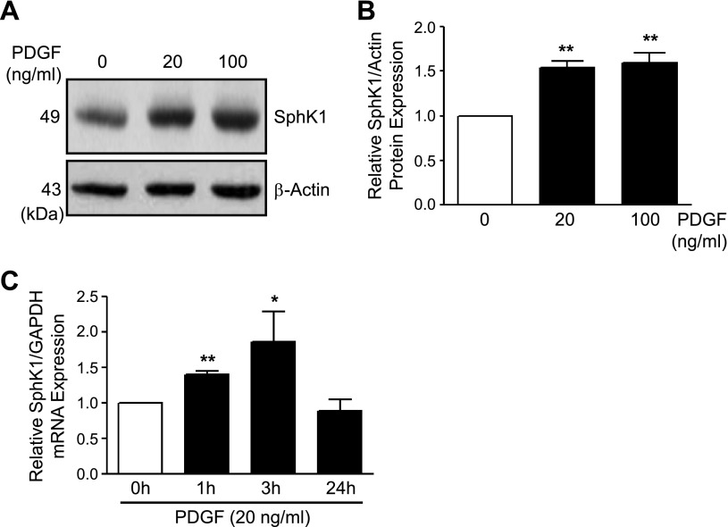
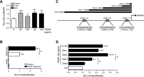
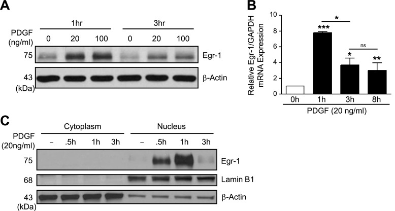
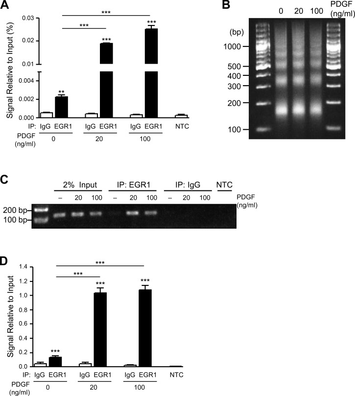
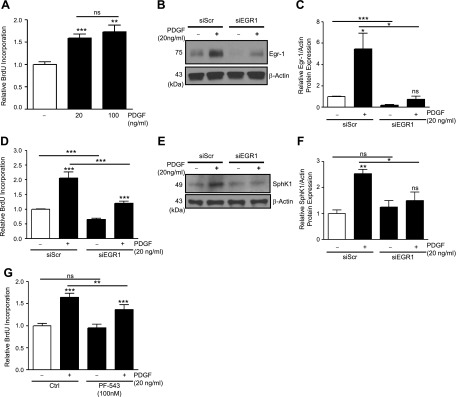
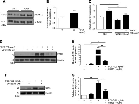
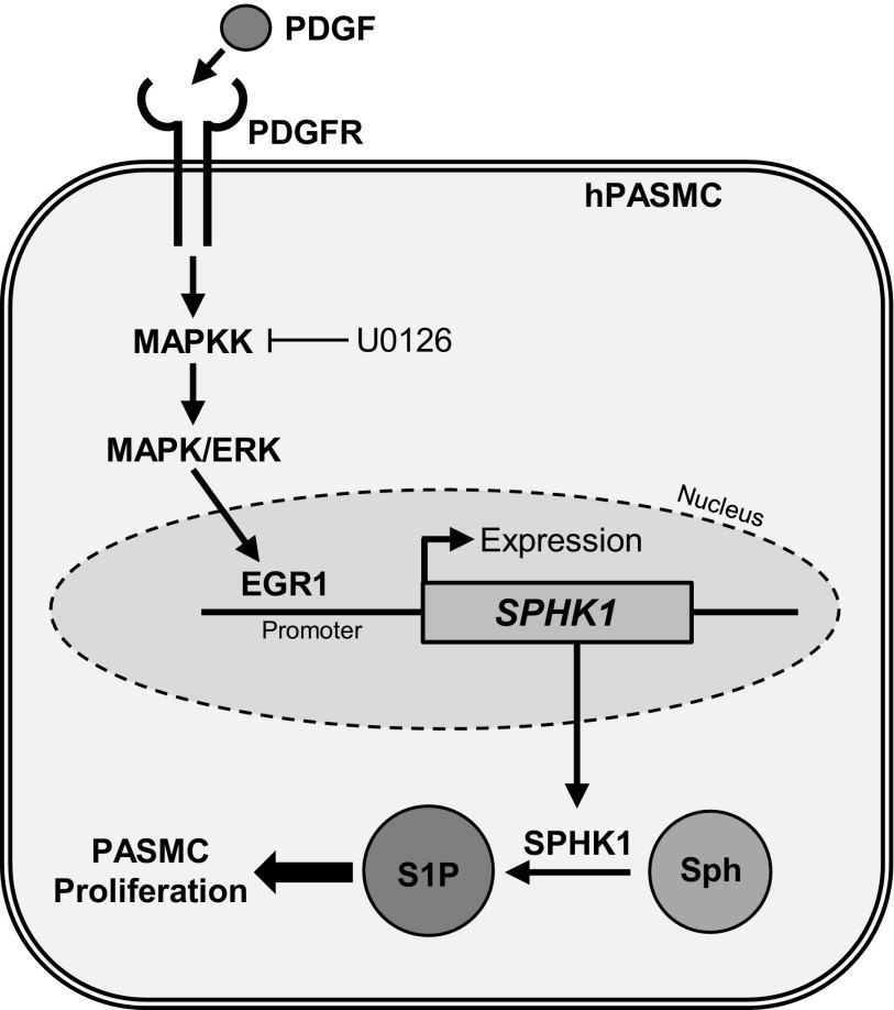
Pulmonary arterial hypertension (PAH) is a progressive, life-threatening disease for which there is currently no curative treatment available. Pathologic changes in this disease involve remodeling of the pulmonary vasculature, including marked proliferation of pulmonary artery smooth muscle cells (PASMCs). Recently, the bioactive lipid sphingosine-1-phosphate (S1P) and its activating kinase, sphingosine kinase 1 (SphK1), have been shown to be upregulated in PAH and promote PASMC proliferation. The mechanisms regulating the transcriptional upregulation of SphK1 in PASMCs are unknown. In this study, we investigated the role of platelet-derived growth factor (PDGF), a PAH-relevant stimuli associated with enhanced PASMC proliferation, on SphK1 expression regulation. In human PASMCs (hPASMCs), PDGF significantly increased SphK1 mRNA and protein expression and induced cell proliferation. Selective inhibition of SphK1 attenuated PDGF-induced hPASMC proliferation. In silico promoter analysis for SphK1 identified several binding sites for early growth response protein 1 (Egr-1), a PDGF-associated transcription factor. Luciferase assays demonstrated that PDGF activates the SphK1 promoter in hPASMCs, and truncation of the 5'-promoter reduced PDGF-induced SphK1 expression. Stimulation of hPASMCs with PDGF induced Egr-1 protein expression, and direct binding of Egr-1 to the SphK1 promoter was confirmed by chromatin immunoprecipitation analysis. Inhibition of ERK signaling prevented induction of Egr-1 by PDGF. Silencing of Egr-1 attenuated PDGF-induced SphK1 expression and hPASMC proliferation. These studies demonstrate that SphK1 is regulated by PDGF in hPASMCs via the transcription factor Egr-1, promoting cell proliferation. This novel mechanism of SphK1 regulation may be a therapeutic target in pulmonary vascular remodeling in PAH.

Keywords: Egr1; PASMC; PDGF; SphK1; gene expression; proliferation.


Figures

Fig. 1.
Fig. 1.
PDGF increases SphK1 expression in hPASMCs. A and B: representative Western blotting images and β-actin-normalized quantification of protein levels demonstrate increased SphK1 expression in hPASMC following stimulation with PDGF-BB (20–100 ng/ml, 6 h). C: SphK1 mRNA expression levels are also increased in hPASMC following PDGF-BB treatment (20 ng/ml, 0–24 h). Results are shown as means ± SE from at least 3 experiments. *P < 0.05, **P < 0.01 vs. untreated control.
Fig. 2.
Fig. 2.
PDGF activates the SphK1 promoter in hPASMCs. A: relative luminescence of secreted Gaussia Luciferase (GLuc) and Secreted Alkaline Phosphatase (SEAP) in a SphK1 promoter dual-reporter system, demonstrating that PDGF (20–100 ng/ml, 1.5 h) stimulates transcriptional activity of the SphK1 promoter (1300 bp) in hPASMCs. B: relative luminescence of a full-length human Sphk1 promoter (∼2.1 kb) cloned into a pGL4 luciferase reporter vector (Firefly/Renilla) demonstrating induction of SphK1 promoter transcriptional activity with PDGF (20 ng/ml, 1.5 h) in hPASMCs. C: representation of the SphK1 promoter containing several putative EGR1 binding sites (EGR1-A, -B, and -C) identified in silico, with black bars depicting SphK1 promoter deletion regions cloned into pGL4 luciferase reporter vectors. D: relative luminescence of Sphk1 promoter deletion constructs demonstrating induction of SphK1 promoter transcriptional activity with PDGF (20 ng/ml, 1.5 h) in hPASMCs. Results are shown as means ± SE from at least 3 experiments. *P < 0.05, **P < 0.01, ***P < 0.001 vs. control unless otherwise indicated.
Fig. 3.
Fig. 3.
PDGF increases EGR1 expression in hPASMCs. A: representative Western blotting images demonstrate increased EGR1 protein expression in human pulmonary artery smooth muscle cells (hPASMC) following stimulation with PDGF-BB (20–100 ng/ml, 1–3 h). B: EGR1 mRNA expression levels are increased in hPASMC following PDGF-BB treatment (20 ng/ml, 0–8 h) relative to GAPDH expression. C: Western blotting with cytoplasmic and nuclear hPASMC fractions demonstrates PDGF-BB treatment (20 ng/ml, 0–3 h) induces EGR1 expression limited to the nucleus, with Lamin B1 used as a nuclear loading control. Results are shown as means ± SE from at least 3 experiments. *P < 0.05, **P < 0.01, ***P < 0.001 vs. control unless otherwise indicated.
Fig. 4.
Fig. 4.
PDGF induces EGR1 binding to the SphK1 promoter in hPASMCs. A: ChIP real-time PCR analysis demonstrates EGR1 binding to the EGR1-“B” site within the SphK1 promoter following stimulation of hPASMC with PDGF (20–100 ng/ml, 1 h), with optimized chromatin digestion of ChIP samples (B). C and D: representative DNA gel electrophoresis of ChIP PCR products demonstrates enhanced EGR1 binding to the EGR1-“B” site within the SphK1 promoter following stimulation of hPASMC with PDGF (20–100 ng/ml, 1 h), with quantification of data signal relative to input. Results are shown as means ± SE from at least 3 experiments. **P < 0.01, ***P < 0.001 vs. IgG control unless otherwise indicated.
Fig. 5.
Fig. 5.
PDGF promotes hPASMC proliferation and SphK1 expression via EGR1. A: BrdU-incorporation assays demonstrate enhanced proliferation of hPASMC following stimulation with PDGF-BB (20–100 ng/ml, 48 h). B and C: siRNA-mediated silencing of EGR1 reduces basal and PDGF-induced (20 ng/ml, 1 h) EGR1 expression, with data quantification. D: siRNA-mediated silencing of EGR1 reduces basal hPASMC proliferation and attenuates PDGF-induced (20 ng/ml, 48 h) proliferation. E and F: siRNA-mediated silencing of EGR1 in hPASMC prevents the induction of SphK1 protein expression by PDGF-BB (20 ng/ml, 6 h), with data quantification. G: selective SphK1 inhibition with PF-543 (100 nM, 1 h pretreatment) attenuates PDGF-induced (20 ng/ml, 48 h) proliferation but not basal proliferation. Results are shown as means ± SE from at least 3 experiments. **P < 0.01, ***P < 0.001 vs. untreated control unless otherwise indicated.
Fig. 6.
Fig. 6.
ERK phosphorylation is required for PDGF-induced EGR1 and SphK1 expression in hPASMC. A and B: representative Western blotting images and ERK-normalized quantification of protein levels demonstrate induction of ERK phosphorylation by PDGF (20 ng/ml, 15 min) in hPASMC. C: U0126-mediated inhibition of ERK phosphorylation reduces basal hPASMC proliferation and attenuates PDGF-induced (20 ng/ml, 48 h) proliferation. D and E: representative Western blotting images and β-actin-normalized quantification of protein levels demonstrate U0126-mediated inhibition of ERK phosphorylation attenuates EGR1 induction by PDGF (20 ng/ml, 1 h) in hPASMC; and attenuates SphK1 induction by PDGF (20 ng/ml, 6 h) in hPASMC (F and G). Results are shown as means ± SE from at least 3 experiments. *P < 0.05, **P < 0.01, ***P < 0.001 vs. untreated control unless otherwise indicated.
Fig. 7.
Fig. 7.
Potential mechanism of PDGF-induced SphK1 expression and proliferation of hPASMCs in pulmonary arterial hypertension (PAH). The PDGF/PDGFR signaling pathway is upregulated in PAH. Activation of this pathway induces phosphorylation of MAPK/ERK, expression and nuclear translocation of the EGR1 transcription factor, and subsequent upregulation of SphK1 gene expression. Increased intracellular SphK1 may then lead to S1P production in hPASMCs, resulting in enhanced proliferation. Abnormal hPASMC proliferation leads to pulmonary vascular remodeling and increased pulmonary vascular resistance in PAH.

References

    1. Agbani EO, Coats P, Mills A, Wadsworth RM. Peroxynitrite stimulates pulmonary artery endothelial and smooth muscle cell proliferation: involvement of ERK and PKC. Pulm Pharmacol Ther 24: 100–109, 2011. - PubMed
    1. Al Husseini A, Bagnato G, Farkas L, Gomez-Arroyo J, Farkas D, Mizuno S, Kraskauskas D, Abbate A, Van Tassel B, Voelkel NF, Bogaard HJ. Thyroid hormone is highly permissive in angioproliferative pulmonary hypertension in rats. Eur Respir J 41: 104–114, 2013. - PubMed
    1. Antoniu SA. Targeting PDGF pathway in pulmonary arterial hypertension. Expert Opin Ther Targets 16: 1055–1063, 2012. - PubMed
    1. Banks MF, Gerasimovskaya EV, Tucker DA, Frid MG, Carpenter TC, Stenmark KR. Egr-1 antisense oligonucleotides inhibit hypoxia-induced proliferation of pulmonary artery adventitial fibroblasts. J Appl Physiol (1985) 98: 732–738, 2005. - PubMed
    1. Berg JT, Breen EC, Fu Z, Mathieu-Costello O, West JB. Alveolar hypoxia increases gene expression of extracellular matrix proteins and platelet-derived growth factor-B in lung parenchyma. Am J Respir Crit Care Med 158: 1920–1928, 1998. - PubMed

MeSH terms

Substances

LinkOut - more resources