

Epigenetic activation of Sox2 gene in the developing vertebrate neural plate
- PMID: 27099369
- PMCID: PMC4907725
- DOI: 10.1091/mbc.E16-01-0042
Epigenetic activation of Sox2 gene in the developing vertebrate neural plate
Abstract
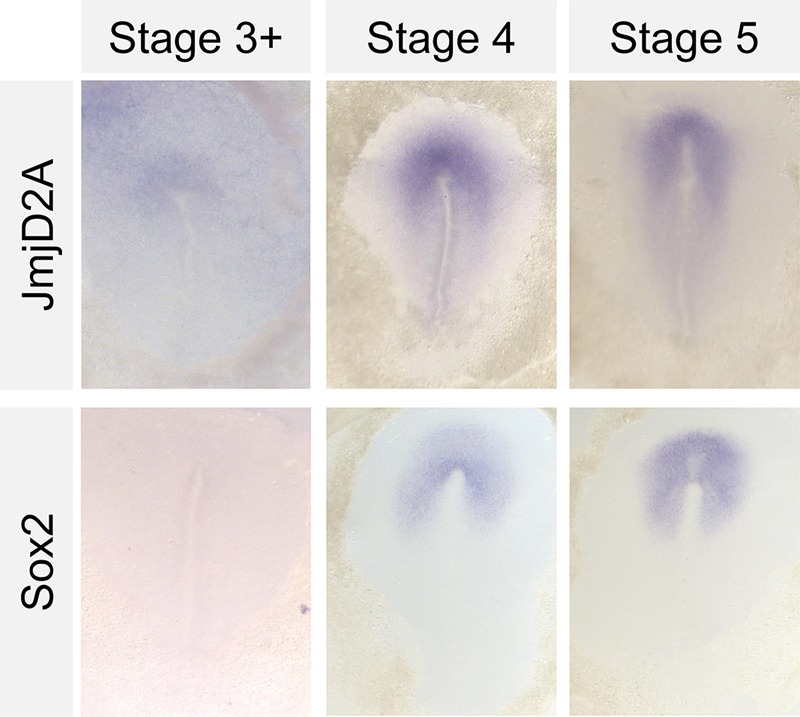
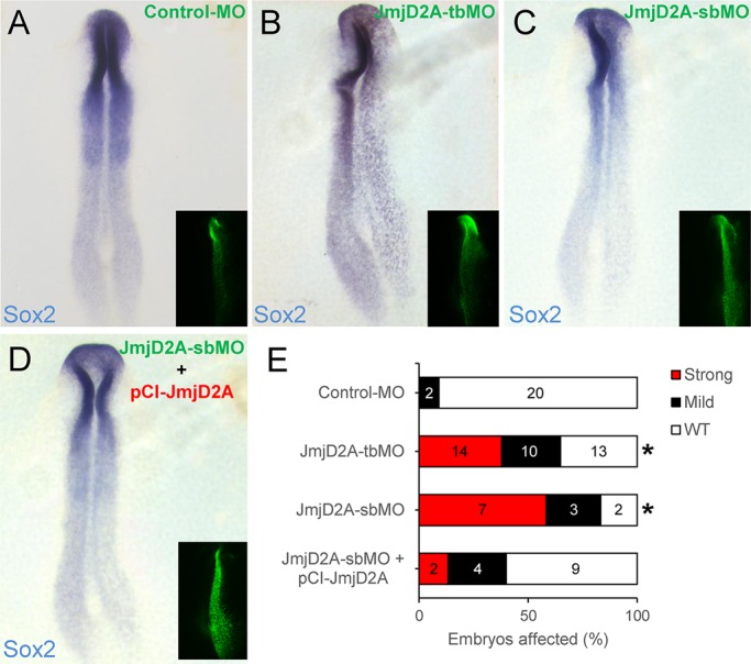
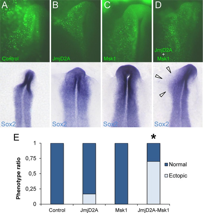
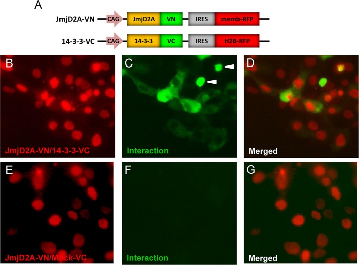
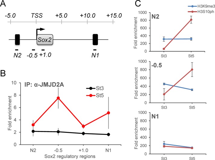
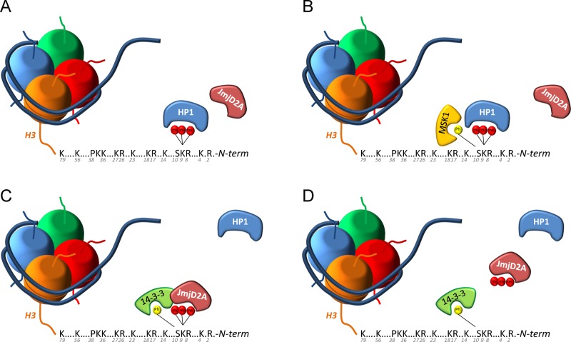
One of the earliest manifestations of neural induction is onset of expression of the neural marker Sox2, mediated by the activation of the enhancers N1 and N2. By using loss and gain of function, we find that Sox2 expression requires the activity of JmjD2A and the Msk1 kinase, which can respectively demethylate the repressive H3K9me3 mark and phosphorylate the activating H3S10 (H3S10ph) mark. Bimolecular fluorescence complementation reveals that the adaptor protein 14-3-3, known to bind to H3S10ph, interacts with JMJD2A and may be involved in its recruitment to regulatory regions of the Sox2 gene. Chromatin immunoprecipitation reveals dynamic binding of JMJD2A to the Sox2 promoter and N-1 enhancer at the time of neural plate induction. Finally, we show a clear temporal antagonism on the occupancy of H3K9me3 and H3S10ph modifications at the promoter of the Sox2 locus before and after the neural plate induction. Taken together, our results propose a series of epigenetic events necessary for the early activation of the Sox2 gene in neural progenitor cells.
© 2016 Bouzas, Marini, et al. This article is distributed by The American Society for Cell Biology under license from the author(s). Two months after publication it is available to the public under an Attribution–Noncommercial–Share Alike 3.0 Unported Creative Commons License (http://creativecommons.org/licenses/by-nc-sa/3.0).
Figures
Similar articles
-
The Pou5f1/Pou3f-dependent but SoxB-independent regulation of conserved enhancer N2 initiates Sox2 expression during epiblast to neural plate stages in vertebrates.Dev Biol. 2011 Apr 15;352(2):354-66. doi: 10.1016/j.ydbio.2010.12.027. Epub 2010 Dec 23. Dev Biol. 2011. PMID: 21185279
-
Tbx6-dependent Sox2 regulation determines neural or mesodermal fate in axial stem cells.Nature. 2011 Feb 17;470(7334):394-8. doi: 10.1038/nature09729. Nature. 2011. PMID: 21331042 Free PMC article.
-
A mechanism regulating the onset of Sox2 expression in the embryonic neural plate.PLoS Biol. 2008 Jan;6(1):e2. doi: 10.1371/journal.pbio.0060002. PLoS Biol. 2008. PMID: 18184035 Free PMC article.
-
Mechanism of cell fate choice between neural and mesodermal development during early embryogenesis.Congenit Anom (Kyoto). 2013 Jun;53(2):61-6. doi: 10.1111/cga.12017. Congenit Anom (Kyoto). 2013. PMID: 23751038 Review.
-
Axial level-dependent molecular and cellular mechanisms underlying the genesis of the embryonic neural plate.Dev Growth Differ. 2016 Jun;58(5):427-36. doi: 10.1111/dgd.12295. Epub 2016 Jun 9. Dev Growth Differ. 2016. PMID: 27279156 Review.
Cited by
-
A catenin-dependent balance between N-cadherin and E-cadherin controls neuroectodermal cell fate choices.Mech Dev. 2018 Aug;152:44-56. doi: 10.1016/j.mod.2018.07.003. Epub 2018 Jul 14. Mech Dev. 2018. PMID: 30009960 Free PMC article.
-
F-box proteins in epigenetic regulation of cancer.Oncotarget. 2017 Nov 16;8(66):110650-110655. doi: 10.18632/oncotarget.22469. eCollection 2017 Dec 15. Oncotarget. 2017. PMID: 29299176 Free PMC article. Review.
-
SOX2 Is a Marker for Stem-like Tumor Cells in Bladder Cancer.Stem Cell Reports. 2017 Aug 8;9(2):429-437. doi: 10.1016/j.stemcr.2017.07.004. Stem Cell Reports. 2017. PMID: 28793245 Free PMC article.
References
-
- Albazerchi A, Stern CD. A role for the hypoblast (AVE) in the initiation of neural induction, independent of its ability to position the primitive streak. Dev Biol. 2007;301:489–503. - PubMed
-
- Bannister AJ, Zegerman P, Partridge JF, Miska EA, Thomas JO, Allshire RC, Kouzarides T. Selective recognition of methylated lysine 9 on histone H3 by the HP1 chromo domain. Nature. 2001;410:120–124. - PubMed
-
- Berger SL. The complex language of chromatin regulation during transcription. Nature. 2007;447:407–412. - PubMed
-
- Bertos NR, Wang AH, Yang XJ. Class II histone deacetylases: structure, function, and regulation. Biochem Cell Biol. 2001;79:243–252. - PubMed
-
- Black JC, Allen A, Van Rechem C, Forbes E, Longworth M, Tschop K, Rinehart C, Quiton J, Walsh R, Smallwood A, et al. Conserved antagonism between JMJD2A/KDM4A and HP1gamma during cell cycle progression. Mol Cell. 2010;40:736–748. - PubMed
Publication types
MeSH terms
Substances
LinkOut - more resources
Full Text Sources
Other Literature Sources
