

Effect of Head and Tongue Posture on the Pharyngeal Airway Dimensions and Morphology in Three-Dimensional Imaging: a Systematic Review
- PMID: 27099695
- PMCID: PMC4837605
- DOI: 10.5037/jomr.2016.7101
Effect of Head and Tongue Posture on the Pharyngeal Airway Dimensions and Morphology in Three-Dimensional Imaging: a Systematic Review
Abstract
Objectives: Natural head position is recommended to be optimal at cone-beam computed tomography acquisition. For standardization purposes in control of treatment outcome, it is clinically relevant to discuss, if a change of posture from natural head position may have an effect on the pharyngeal airway dimensions and morphology, during computed tomography, cone-beam computed tomography or magnetic resonance imaging acquisition. This was the aim of the present literature review study for purposes of valid evidence, which was hypothesized, to be present.
Material and methods: This systematic literature review has been registered in PROSPERO database with following number: CRD42015024567. A systematic literature search performed in PubMed, Embase and Cochrane was carried out in order to evaluate if the effect of human head or tongue posture has an effect on upper airway dimensions and morphology in CT, CBCT or MRI. Study quality assessment was performed. Predictor variable was head and tongue posture. Endpoints were numerical values of upper airway dimensions and morphology.
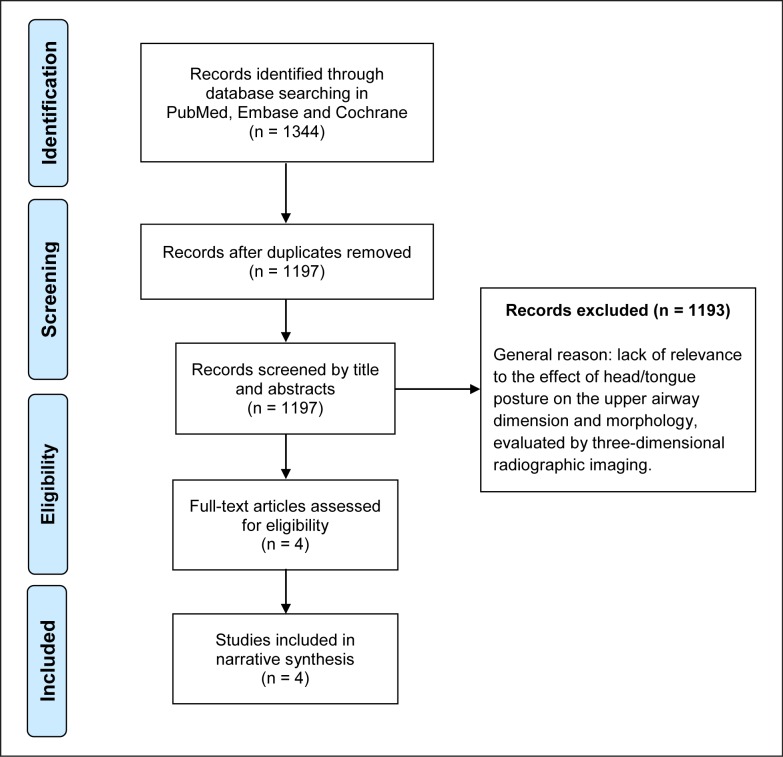
Results: Overall 1344 articles (Embase 1063, PubMed 269, and Cochrane 12) resulted in four included publications. Quality assessments revealed poor quality and low-level evidence by 46 - 67% of the maximum achievable score. Heterogeneous methodology made a meta-analysis impossible, consequently a narrative synthesis was performed.
Conclusions: Limited, poor quality and low evidence level literature is available on the effect of head posture on upper airway dimensions and morphology in three-dimensional imaging. Valid evidence requires a standardized method of head and tongue posture during image acquisition in future studies.
Keywords: cone-beam computed tomography; magnetic resonance imaging; obstructive sleep apnea; orthognathic surgery; posture.
Figures
Similar articles
-
Pharyngeal Airway Dimensions and Head Posture in Obstructive Sleep Apnea Patients with and without Morphological Deviations in the Upper Cervical Spine.J Oral Maxillofac Res. 2017 Sep 30;8(3):e4. doi: 10.5037/jomr.2017.8304. eCollection 2017 Jul-Sep. J Oral Maxillofac Res. 2017. PMID: 29142656 Free PMC article.
-
The effect of altered head and tongue posture on upper airway volume based on a validated upper airway analysis-An MRI pilot study.Orthod Craniofac Res. 2020 Feb;23(1):102-109. doi: 10.1111/ocr.12348. Epub 2019 Nov 20. Orthod Craniofac Res. 2020. PMID: 31550076
-
Does Head and Neck Posture Affect Cone-Beam Computed Tomography Assessment of the Upper Airway?J Oral Maxillofac Surg. 2023 Jun;81(6):721-733. doi: 10.1016/j.joms.2023.01.016. Epub 2023 Feb 23. J Oral Maxillofac Surg. 2023. PMID: 36841260
-
Impact of Bimaxillary Advancement Surgery on the Upper Airway and on Obstructive Sleep Apnea Syndrome: a Meta-Analysis.Sci Rep. 2018 Apr 10;8(1):5756. doi: 10.1038/s41598-018-24142-3. Sci Rep. 2018. PMID: 29636515 Free PMC article.
-
Posterior Airway Changes Following Orthognathic Surgery in Obstructive Sleep Apnea.J Oral Maxillofac Surg. 2018 May;76(5):1093.e1-1093.e21. doi: 10.1016/j.joms.2017.11.035. Epub 2017 Dec 6. J Oral Maxillofac Surg. 2018. PMID: 29288649
Cited by
-
Three-dimensional assessment of Upper Airway in Class III patients with different facial patterns.J Clin Exp Dent. 2023 Oct 1;15(10):e821-e826. doi: 10.4317/jced.60856. eCollection 2023 Oct. J Clin Exp Dent. 2023. PMID: 37933396 Free PMC article.
-
Cone beam computed tomography in assessment on pharynx effects of orthopedic-surgical treatment - a review of the literature.Sleep Sci. 2019 Apr-Jun;12(2):106-109. doi: 10.5935/1984-0063.20190066. Sleep Sci. 2019. PMID: 31879543 Free PMC article. Review.
-
Pharyngeal Airway Dimensions and Head Posture in Obstructive Sleep Apnea Patients with and without Morphological Deviations in the Upper Cervical Spine.J Oral Maxillofac Res. 2017 Sep 30;8(3):e4. doi: 10.5037/jomr.2017.8304. eCollection 2017 Jul-Sep. J Oral Maxillofac Res. 2017. PMID: 29142656 Free PMC article.
-
Effects of 3D Airway Geometry on the Airflow of Adults with Cleft Lip and Palate and Obstructive Sleep Apnea: A Functional Imaging Study.Sleep Sci. 2023 Nov 22;16(4):e430-e438. doi: 10.1055/s-0043-1776868. eCollection 2023 Dec. Sleep Sci. 2023. PMID: 38197022 Free PMC article.
-
The use of CBCT in orthodontics with special focus on upper airway analysis in patients with sleep-disordered breathing.Dentomaxillofac Radiol. 2024 Mar 25;53(3):178-188. doi: 10.1093/dmfr/twae001. Dentomaxillofac Radiol. 2024. PMID: 38265247 Free PMC article.
References
Publication types
LinkOut - more resources
Full Text Sources
Other Literature Sources