. 2016 Jan-Mar;8(1):21-33.

MicroRNAs: The Role in Autoimmune Inflammation

Affiliations

MicroRNAs: The Role in Autoimmune Inflammation

N M Baulina et al. Acta Naturae. 2016 Jan-Mar.

Abstract

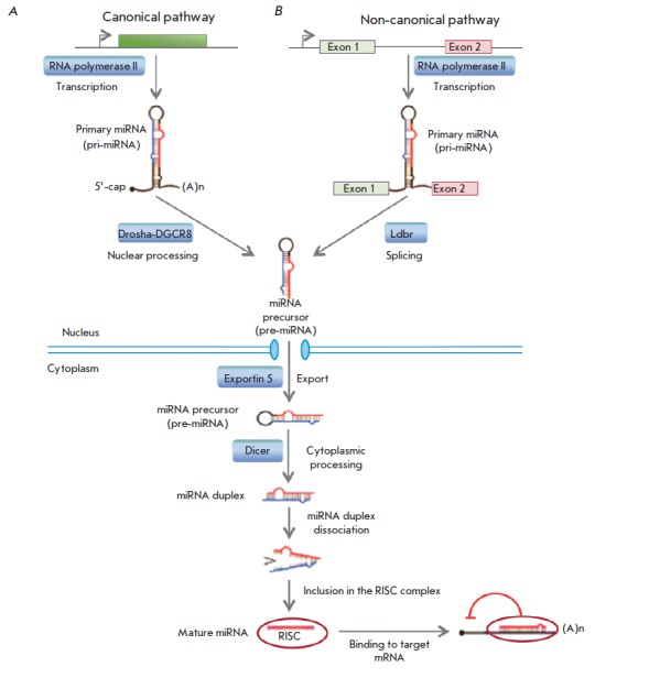
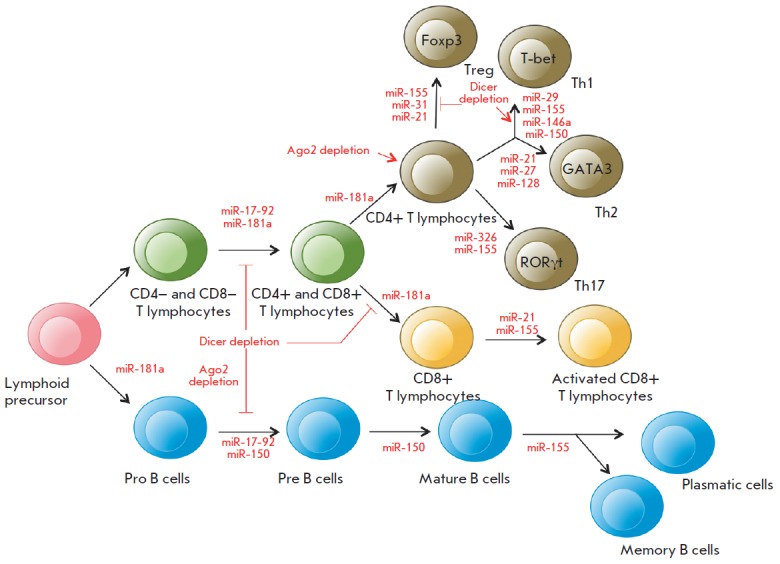
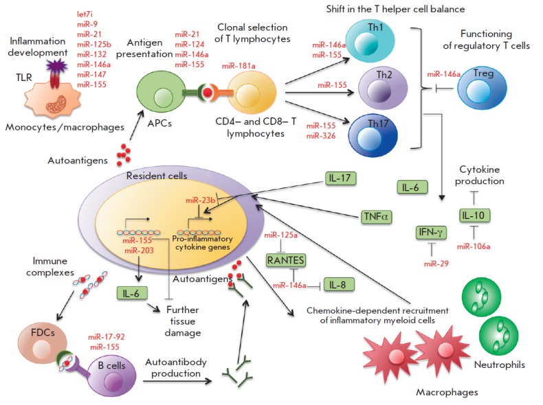
MicroRNAs (miRNAs) are small non-coding RNA molecules that regulate gene expression at the post-transcriptional level through base-pairing predominantly with a 3'-untranslated region of target mRNA, followed by mRNA degradation or translational repression. Totally, miRNAs change, through a complex regulatory network, the expression of more than 60% of human genes. MiRNAs are key regulators of the immune response that affect maturation, proliferation, differentiation, and activation of immune cells, as well as antibody secretion and release of inflammatory mediators. Disruption of this regulation may lead to the development of various pathological conditions, including autoimmune inflammation. This review summarizes the data on biogenesis and the mechanisms of miRNA action. We discuss the role of miRNAs in the development and the action of the immune system, as well as in the development of an autoimmune inflammatory response. Special attention is given to the role of miRNAs in the autoimmune inflammation in multiple sclerosis, which is a serious socially significant disease of the central nervous system. Currently, a lot of research is focused on this problem.

Keywords: autoimmune inflammation; microRNA; multiple sclerosis.


Figures

Fig. 1
Fig. 1
MiRNA biogenesis. A. The canonical pre-miRNA pathway produces pre-miRNAs through cleavage of pri-miRNA transcripts by the Drosha-DGCR8 microprocessor complex. B. The non-canonical pathway. Mirtrons are spliced and debranched by the Ldbr enzyme, after which they fold into pre-miRNA hairpins. Then, the pathways merge. The green box indicates a miRNA gene; exons 1 and 2 are exons of the host gene encoding intronic miRNA.
Fig. 2
Fig. 2
Redundancy and pleiotropy of the miRNA regulatory system. The STAT3 gene (green oval) encodes a transcriptional factor. Red ovals are miRNAs downregulating the STAT3 gene expression. Each of the miRNAs inhibits expression of other target mRNAs (blue, violet, and light blue ovals). Additional miRNAs that may affect the STAT3 gene expression are listed on the right-hand side. The regulatory network was simulated using the Mirtarbase database (http://mirtarbase.mbc.nctu.edu.tw/index.php).
Fig. 3
Fig. 3
The role of miRNAs in the differentiation of T and B cells (modified from [28]). Th1, Th2, and Th17 are T helper cells; Treg are regulatory T cells; Foxp3, T-bet, GATA3, and RORγt are transcription factors required for the normal development of various T helper cell subsets. See the text for details.
Fig. 4
Fig. 4
The role of miRNAs in autoimmune inflammation (modified from [35]). APCs are antigen-presenting cells; Th1, Th2, and Th17 are T helper cells; Treg are regulatory T cells; FDCs are follicular dendritic cells. See text for more details.

References

    1. Rebane A., Akdis C.A.. J. Allergy Clin. Immunol. 2013;132:15–26. - PubMed
    1. Reinhart B.J., Slack F.J., Basson M., Pasquinelli A.E., Bettinger J.C., Rougvie A.E., Horvitz H.R., Ruvkun G.. Nature. 2000;403:901–906. - PubMed
    1. Kamanu T.K., Radovanovic A., Archer J.A., Bajic V.B.. Sci. Rep. 2013;3:2940. - PMC - PubMed
    1. Eulalio A., Mano M.. J. Biomol. Screen. 2015;20:1003–1017. - PubMed
    1. Meunier J., Lemoine F., Soumillon M., Liechti A., Weier M., Guschanski K., Hu H., Khaitovich P., Kaessmann H.. Genome Res. 2013;23:34–45. - PMC - PubMed

LinkOut - more resources