

Marmosets: A Neuroscientific Model of Human Social Behavior
- PMID: 27100195
- PMCID: PMC4840471
- DOI: 10.1016/j.neuron.2016.03.018
Marmosets: A Neuroscientific Model of Human Social Behavior
Abstract
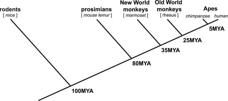
The common marmoset (Callithrix jacchus) has garnered interest recently as a powerful model for the future of neuroscience research. Much of this excitement has centered on the species' reproductive biology and compatibility with gene editing techniques, which together have provided a path for transgenic marmosets to contribute to the study of disease as well as basic brain mechanisms. In step with technical advances is the need to establish experimental paradigms that optimally tap into the marmosets' behavioral and cognitive capacities. While conditioned task performance of a marmoset can compare unfavorably with rhesus monkey performance on conventional testing paradigms, marmosets' social behavior and cognition are more similar to that of humans. For example, marmosets are among only a handful of primates that, like humans, routinely pair bond and care cooperatively for their young. They are also notably pro-social and exhibit social cognitive abilities, such as imitation, that are rare outside of the Apes. In this Primer, we describe key facets of marmoset natural social behavior and demonstrate that emerging behavioral paradigms are well suited to isolate components of marmoset cognition that are highly relevant to humans. These approaches generally embrace natural behavior, which has been rare in conventional primate testing, and thus allow for a new consideration of neural mechanisms underlying primate social cognition and signaling. We anticipate that through parallel technical and paradigmatic advances, marmosets will become an essential model of human social behavior, including its dysfunction in neuropsychiatric disorders.
Copyright © 2016 Elsevier Inc. All rights reserved.
Figures
References
-
- Abbott DH. Behavioral and physiological suppression of fertility in subordinate marmoset monkeys. American Journal of Primatology. 1984;6:169–186. - PubMed
-
- Abbott DH, Saltzman W, Schutz-Darken NJ, Smith TE. Specific neuroendocrine mechanisms not involving generalized stress mediate social regulation of fremale reproduction in cooperative breeding marmoset monkeys. In: Carver CS, Kirkpatrick B, Lederhendler LL, editors. The Integrative Neurobiology of Affiliation. Annals of the New York Academy of Sciences; NY: 1997. pp. 219–238. - PubMed
Publication types
MeSH terms
Grants and funding
LinkOut - more resources
Full Text Sources
Other Literature Sources
Miscellaneous
