

Lupus Panniculitis as an Initial Manifestation of Systemic Lupus Erythematosus: A Case Report
- PMID: 27100438
- PMCID: PMC4845842
- DOI: 10.1097/MD.0000000000003429
Lupus Panniculitis as an Initial Manifestation of Systemic Lupus Erythematosus: A Case Report
Abstract
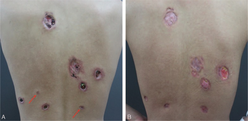
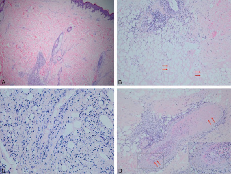
Lupus erythematosus panniculitis (LEP) is a variant of chronic cutaneous lupus erythematosus (CCLE). Reported cases of LEP lesions before the diagnosis of systemic lupus erythematosus (SLE) were very rare; only 9 cases have been reported, to the best of our knowledge. We now describe the case of a 19-year-old male patient, with an overall review of the English literature. In the earliest stage of the present case, nodules and ulcers involved his left leg and face, with no other accompanied symptoms. The skin lesions disappeared after treatment with methylprednisolone, 16 mg/d for 1 month. Seven months after discontinuing methylprednisolone, the cutaneous nodules and ulcers on his back recurred and were accompanied by fever, hair loss, and polyarthritis. Blood tests revealed leucopenia, positive antinuclear antibody and Smith antibody, and proteinuria. Histopathological findings were most consistent with LEP. This was followed sequentially by the diagnosis of SLE. The patient improved again after treatment with methylprednisolone and cyclophosphamide.Patients with LEP should have regular follow-ups because the development of SLE is possible. Early diagnosis and proper treatment is pivotal to improve the prognosis of such patients.
Conflict of interest statement
The authors have no conflicts of interest to disclose.
Figures
Similar articles
-
Lupus erythematosus panniculitis in a 10-year-old female child with severe systemic lupus erythematosus: A case report.Medicine (Baltimore). 2018 Jan;97(3):e9571. doi: 10.1097/MD.0000000000009571. Medicine (Baltimore). 2018. PMID: 29504978 Free PMC article.
-
Atypical lupus erythematosus panniculitis progressing to antinuclear antibody-negative systemic lupus erythematosus.J Cutan Med Surg. 2012 Sep-Oct;16(5):361-4. doi: 10.1177/120347541201600516. J Cutan Med Surg. 2012. PMID: 22971314
-
[Characteristics of the course of lupus erythematosus panniculitis in a retrospective analysis of 17 patients].Orv Hetil. 2023 Feb 5;164(5):172-178. doi: 10.1556/650.2023.32692. Print 2023 Feb 5. Orv Hetil. 2023. PMID: 36739549 Hungarian.
-
[A case of systemic lupus erythematosus complicated with psoriasis vulgaris].Nihon Rinsho Meneki Gakkai Kaishi. 2003 Dec;26(6):341-5. doi: 10.2177/jsci.26.341. Nihon Rinsho Meneki Gakkai Kaishi. 2003. PMID: 14752935 Review. Japanese.
-
A case of toxic epidermal necrolysis-like skin lesions with systemic lupus erythematosus and review of the literature.Lupus. 2013 Jul;22(8):839-46. doi: 10.1177/0961203313492242. Epub 2013 Jun 11. Lupus. 2013. PMID: 23761100 Review.
Cited by
-
Lupus profundus limited to a site of trauma: Case report and review of the literature.Int J Womens Dermatol. 2017 May 5;3(2):117-120. doi: 10.1016/j.ijwd.2017.03.002. eCollection 2017 Jun. Int J Womens Dermatol. 2017. PMID: 28560307 Free PMC article.
-
Sustained Drug Free Remission of Lupus Panniculitis with Methotrexate: A Case Report From Nepal.JNMA J Nepal Med Assoc. 2018 Nov-Dec;56(214):963-966. doi: 10.31729/jnma.3885. JNMA J Nepal Med Assoc. 2018. PMID: 31065144 Free PMC article.
-
Lupus Profundus: A Case Report from Pakistan.Cureus. 2018 May 28;10(5):e2697. doi: 10.7759/cureus.2697. Cureus. 2018. PMID: 30062071 Free PMC article.
-
Lupus panniculitis as the initial presentation of systemic lupus erythematosus triggered by COVID-19 infection: case report and literature review.Oxf Med Case Reports. 2023 Nov 28;2023(11):omad129. doi: 10.1093/omcr/omad129. eCollection 2023 Nov. Oxf Med Case Reports. 2023. PMID: 38033407 Free PMC article.
-
Purulent lupus panniculitis unmasked by FDG-PET/CT scan: A case report.Medicine (Baltimore). 2016 Nov;95(48):e5478. doi: 10.1097/MD.0000000000005478. Medicine (Baltimore). 2016. PMID: 27902603 Free PMC article.
References
-
- Arai S, Katsuoka K. Clinical entity of Lupus erythematosus panniculitis/lupus erythematosus profundus. Autoimmun Rev 2009; 8:449–452. - PubMed
-
- Park HS, Choi JW, Kim BK, et al. Lupus erythematosus panniculitis: clinicopathological, immunophenotypic, and molecular studies. Am J Dermatopathol 2010; 32:24–30. - PubMed
-
- Ng PP, Tan SH, Tan T. Lupus erythematosus panniculitis: a clinicopathologic study. Int J Dermatol 2002; 41:488–490. - PubMed
-
- Martens PB, Moder KG, Ahmed I. Lupus panniculitis: clinical perspectives from a case series. J Rheumatol 1999; 26:68–72. - PubMed
-
- Diaz-Jouanen E, DeHoratius RJ, Alarcon-Segovia D, et al. Systemic lupus erythematosus presenting as panniculitis (lupus profundus). Ann Intern Mes 1975; 82:376–379. - PubMed
Publication types
MeSH terms
LinkOut - more resources
Full Text Sources
Other Literature Sources
Medical
Miscellaneous
