. 2016 Apr 21;11(4):e0153402.
doi: 10.1371/journal.pone.0153402. eCollection 2016.

M-Cells Contribute to the Entry of an Oral Vaccine but Are Not Essential for the Subsequent Induction of Protective Immunity against Francisella tularensis

Affiliations

M-Cells Contribute to the Entry of an Oral Vaccine but Are Not Essential for the Subsequent Induction of Protective Immunity against Francisella tularensis

Aimee L Cunningham et al. PLoS One. .

Abstract

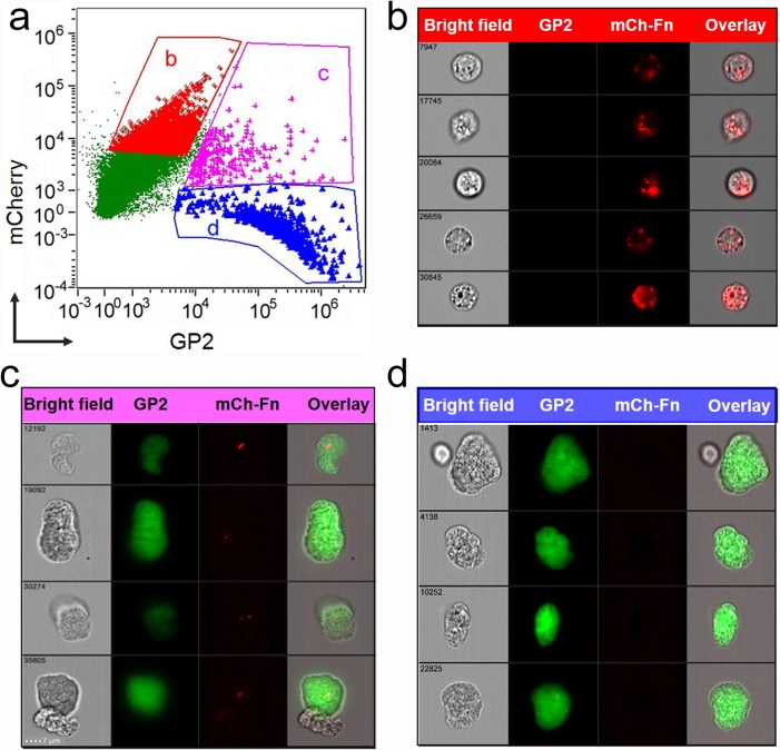
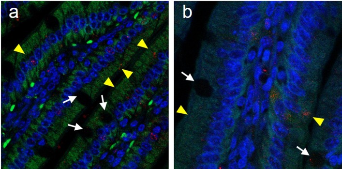
M-cells (microfold cells) are thought to be a primary conduit of intestinal antigen trafficking. Using an established neutralizing anti-RANKL (Receptor Activator of NF-κB Ligand) antibody treatment to transiently deplete M-cells in vivo, we sought to determine whether intestinal M-cells were required for the effective induction of protective immunity following oral vaccination with ΔiglB (a defined live attenuated Francisella novicida mutant). M-cell depleted, ΔiglB-vaccinated mice exhibited increased (but not significant) morbidity and mortality following a subsequent homotypic or heterotypic pulmonary F. tularensis challenge. No significant differences in splenic IFN-γ, IL-2, or IL-17 or serum antibody (IgG1, IgG2a, IgA) production were observed compared to non-depleted, ΔiglB-vaccinated animals suggesting complementary mechanisms for ΔiglB entry. Thus, we examined other possible routes of gastrointestinal antigen sampling following oral vaccination and found that ΔiglB co-localized to villus goblet cells and enterocytes. These results provide insight into the role of M-cells and complementary pathways in intestinal antigen trafficking that may be involved in the generation of optimal immunity following oral vaccination.


Conflict of interest statement

Competing Interests: The authors have declared that no competing interests exist.

Figures

Fig 1
Fig 1. Uptake of ΔiglB vaccine strain by intestinal M-cells following oral inoculation.
BALB/c mice (4–6 wks) were orally vaccinated with mCherry-expressing ΔiglB (108 CFU/100 μL). Small intestines were collected at 90 min. post-vaccination and Peyer’s patches were excised to generate single cell suspensions. Cells were labeled with AF-488 conjugated α-GP2 antibody and subjected to cytometry imaging analysis. (a) Dot-plot depicts GP2 and mCherry intensity of each examined cell and three gated cell populations: (b) mCherryhiGP2low, (c) mCherryhiGP2hi, and (d) mCherrylowGP2hi. The representative cell images of these three gated populations are shown in (b) ΔiglB residing in non-M-cells, (c) ΔiglB residing in M-cells, and (d) M-cells with no ΔiglB, respectively. Representative images from 2 independent experiments are shown.
Fig 2
Fig 2. M-cells are depleted with αRANKL antibody treatment.
BALB/c mice (n = 3 per group) were treated i.p. with 250 μg of either rat Ig (a-b) or αRANKL antibody IK22-5 (c-d) on days 0, 2, 4, and 6. On day 8 (a-c) or day 16 (d), animals were sacrificed and Peyer's patches (PP) collected and stained with rhodamine-labeled UEA-1 for whole mount imaging. Representative images showing normal morphology of intact (ratIg-treated) PP were taken using combined phase contrast and fluorescence at 40x (a), or labeled M cells (480 in rat Ig-treated PP on day 8, 65 M-cells in αRANKL-treated PP on day 8, or 476 in αRANKL-treated PP on day 16) all with fluorescence alone at 100x (b-d). M-cells in PP whole mounts were quantified for naive, rat Ig-treated, and αRANKL-treated groups (e), with αRANKL treatment inducing a significant decrease in M-cells (***p < 0.001) compared to naive or rat Ig-treated animals at day 8 or αRANKL-treated animals upon PP repopulation (day 16). Representative images from 3 experiments are shown.
Fig 3
Fig 3. Depletion of M-cells does not affect ability to survive subsequent pulmonary challenge or mount an immune response.
BALB/c mice (n = 10 per group for a & b, n = 3 per group for c & d) were treated i.p. with rat Ig or αRANKL antibody (250 μg) on days 0, 2, 4, and 6, and then, along with an untreated group of mice (indicated as ΔiglB), were vaccinated orally with ΔiglB (1000 CFU for a & b, 105 CFU for c & d) on day 8. Naïve mice receiving PBS orally were used as the non-vaccinated control. (a-b) After 30 days, all animals were intranasally challenged with 45,000 CFU of LVS (~10 LD50) and were monitored daily for weight changes (a) and survival (b). Representative results from 3 experiments are shown. (c-d) After a resting period of 21 days to allow for clearance of the vaccine, mice were bled to obtain sera, and then sacrificed for spleen collection. (c) ELISpots for IFN-γ, IL-2, and IL-17 were performed using single-cell splenocytes stimulated with 1 μg of UV-killed ΔiglB. Spot numbers of 3 individual spleens with triplicate, 9 for each group, are shown with mean ± standard error. Representative of 2 independent experiments. (d) ELISAs were conducted to determine serum antibody responses (total antibody, IgG1, and IgG2a) to UV-killed ΔiglB. Representative results from 3 experiments are shown. * p <0.05, *** p < 0.001.
Fig 4
Fig 4. Goblet cells can take up Francisella tularensis by 90 minutes after oral vaccination.
BALB/c mice (n = 3) were orally vaccinated with mCherry-ΔiglB (approximately 108 CFU) and rested for 90 minutes prior to sacrifice for collection of whole intestines, which were paraffin embedded, sectioned, and stained for (a) periodic acid Schiff to visualize GCs (black arrowheads) and (b-c) confocal analysis with nuclear stain DAPI (blue), mucin marker anti-MUC-2 (green), and GC surface marker anti-cytokeratin-18 (pink), with GCs shown by white arrowheads. (c) High magnification of a goblet cell (circled), stained with both MUC-2 and cytokeratin-18, which has taken up ΔiglB (red). (d) Single cells were prepared from intestines of similarly vaccinated mice, labeled with FITC-anti-MUC-2 (green), and subjected to cytometry imaging to visualize the uptake of mCherry-ΔiglB (red) by goblet cells. BF, bright field. Representative images from 2 separate experiments are shown.
Fig 5
Fig 5. Uptake of mCherry-ΔiglB by enterocytes.
BALB/c mice (n = 3) were orally vaccinated with mCherry-ΔiglB (approximately 108 CFU) and rested for 90 minutes prior to sacrifice for collection of whole intestines, which were paraffin embedded, sectioned, and stained for confocal analysis with nuclear stain DAPI (blue), and with mCherry-ΔiglB in GCs shown by white arrows and mCherry-ΔiglB in enterocytes shown by yellow arrowheads. (a) 630x and (b) 1000x.

References

    1. Brandtzaeg P, Baekkevold ES, Morton HC. From B to A the mucosal way. Nat Immunol. 2001;2(12):1093–4. Epub 2001/11/29. 10.1038/ni1201-1093 . - DOI - PubMed
    1. Corr SC, Gahan CC, Hill C. M-cells: origin, morphology and role in mucosal immunity and microbial pathogenesis. FEMS Immunol Med Microbiol. 2008;52(1):2–12. Epub 2007/12/18. 10.1111/j.1574-695X.2007.00359.x . - DOI - PubMed
    1. Ray HJ, Cong Y, Murthy AK, Selby DM, Klose KE, Barker JR, et al. Oral live vaccine strain-induced protective immunity against pulmonary Francisella tularensis challenge is mediated by CD4+ T cells and antibodies, including immunoglobulin A. Clin Vaccine Immunol. 2009;16(4):444–52. Epub 2009/02/13. 10.1128/CVI.00405-08 - DOI - PMC - PubMed
    1. Dietrich G, Griot-Wenk M, Metcalfe IC, Lang AB, Viret JF. Experience with registered mucosal vaccines. Vaccine. 2003;21(7–8):678–83. Epub 2003/01/18. doi: S0264410X02005790 [pii]. . - PubMed
    1. Levine MM. Can needle-free administration of vaccines become the norm in global immunization? Nat Med. 2003;9(1):99–103. Epub 2003/01/07. 10.1038/nm0103-99 . - DOI - PubMed

Publication types

LinkOut - more resources