. 2016 Apr 21;11(4):e0152996.
doi: 10.1371/journal.pone.0152996. eCollection 2016.

CIP2A Promotes T-Cell Activation and Immune Response to Listeria monocytogenes Infection

Affiliations

CIP2A Promotes T-Cell Activation and Immune Response to Listeria monocytogenes Infection

Christophe Côme et al. PLoS One. .

Abstract

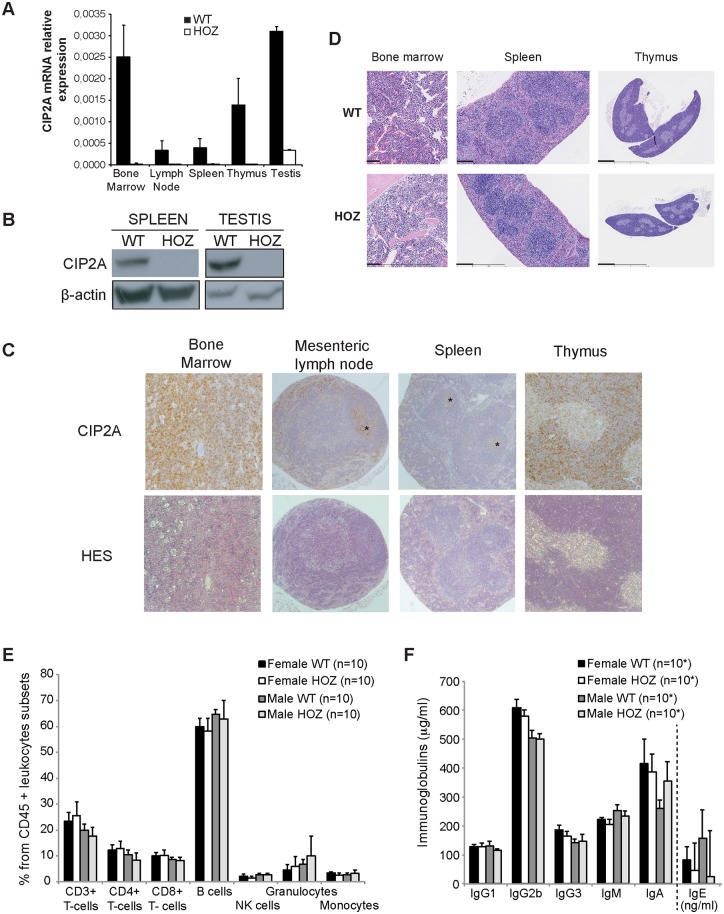
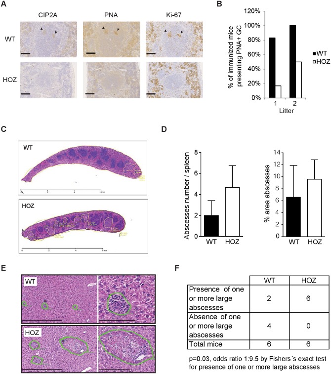
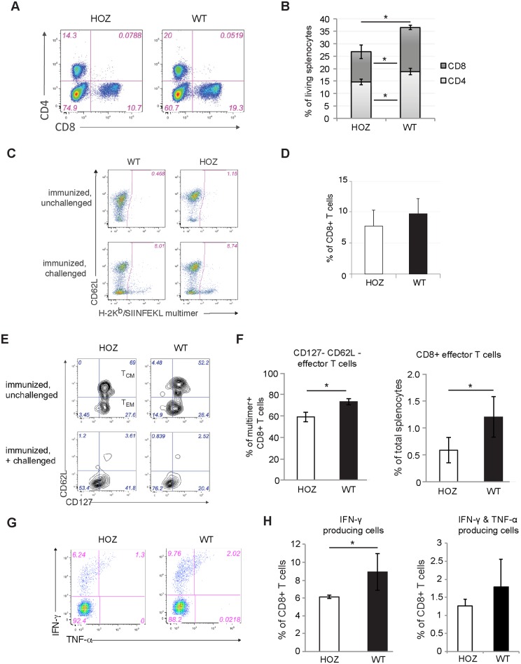
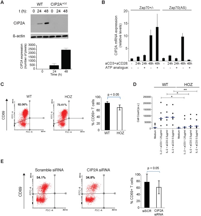
The oncoprotein Cancerous Inhibitor of Protein Phosphatase 2A (CIP2A) is overexpressed in most malignancies and is an obvious candidate target protein for future cancer therapies. However, the physiological importance of CIP2A-mediated PP2A inhibition is largely unknown. As PP2A regulates immune responses, we investigated the role of CIP2A in normal immune system development and during immune response in vivo. We show that CIP2A-deficient mice (CIP2AHOZ) present a normal immune system development and function in unchallenged conditions. However when challenged with Listeria monocytogenes, CIP2AHOZ mice display an impaired adaptive immune response that is combined with decreased frequency of both CD4+ T-cells and CD8+ effector T-cells. Importantly, the cell autonomous effect of CIP2A deficiency for T-cell activation was confirmed. Induction of CIP2A expression during T-cell activation was dependent on Zap70 activity. Thus, we reveal CIP2A as a hitherto unrecognized mediator of T-cell activation during adaptive immune response. These results also reveal CIP2AHOZ as a possible novel mouse model for studying the role of PP2A activity in immune regulation. On the other hand, the results also indicate that CIP2A targeting cancer therapies would not cause serious immunological side-effects.


Conflict of interest statement

Competing Interests: The authors have declared that no competing interests exist.

Figures

Fig 1
Fig 1. Expression of CIP2A by murine lymphoid organs does not impact the immune system of unchallenged CIP2AHOZ animals.
(A) Semi-quantitative RT-PCR for CIP2A RNA in main lymphoid organs and testis from WT and CIP2AHOZ adult mice. Average expression from at least 3 mice (despite testis; S1 Table). (B) Protein expression of spleen and testis from WT and CIP2AHOZ adult mice. Mouse beta-actin was used for normalization. (C) CIP2A-IHC and hematoxilin-eosin staining of common lymphoid organs from adult wild-type mice. Asterix indicate CIP2A expression in germinal centers of lymph nodes and spleen. (D) Hematoxilin and eosin immunohistological staining reveals normal morphology of lymphoid tissues in control and CIP2AHOZ mice. Thick scale bar: 100 μm (Bone marrow); 250 μm (Spleen); 1 mm (Thymus). (E) Flow cytometry analysis of frequencies of leukocyte subpopulations in peripheral blood of WT or CIP2AHOZ females and males (10 mice per condition). (F) Principal immunoglobulins content (μg/ml; except for IgE, ng/ml) in the peripheral blood of WT or CIP2AHOZ adult mice (*: 10 mice per condition, except for IgE, with samples from 9 WT or CIP2AHOZ females, 9 WT males and 7 CIP2AHOZ males). (E, F) Medians are represented and p-values calculated based on a Wilcoxon rank-sum test.
Fig 2
Fig 2. Impaired adaptive immune response in CIP2A deficient mice.
(A) Immunohistochemical staining for CIP2A, germinal center marker peanut agglutinin (PNA), and proliferation marker Ki-67 in spleen from immunized WT and CIP2AHOZ mice. Arrows indicate positive staining of germinal center of indicated antigens in WT tissues. Scale bar represents 100 μm. (B) Percentage of immunized mice presenting a PNA positive staining in germinal center from two independent mouse cohorts of 6 WT and 6 CIP2AHOZ females, and 7 WT and 6 CIP2AHOZ females respectively. (C) Haematoxilin histological staining of spleen after primary infection with L.m. Bacterial abscesses are highlighted in yellow. Scale bar: 6 mm. (D) Abscesses number and percentage of the abscesses area compared to spleen total area after L.m. primary infection. Average of 2 WT and 3 CIP2AHOZ females is shown. Mann-Whitney t test. (E) Hematoxilin-eosin histological staining of liver presenting inflammatory lesions developed in response to second infection with intracellular bacteria L.m (5 days post recall infection). The right panels present higher amplifications of the lesions on the left. Scale bar represents 500 μm (left panel) or 100 μm (right panel). Representative pictures of 6 WT and 6 CIP2AHOZ females analyzed. (F) Quantitative and statistical analysis of the number of large abscesses (> 10 μm2) as described in (E).
Fig 3
Fig 3. Impaired T-cell activation in CIP2AHOZ mice in vivo.
(A-H) Flow cytometry analysis of splenocytes from 3 WT and 4 CIP2AHOZ mice five days after recall infection with high-dose L.m.-OVA. (A) Representative flow cytometry analysis for CD4+ and CD8+ T cells. (B) Percentage of CD4+ and CD8+ splenocytes from analysis described in (A). * p < 0.05, Two-tailed T-test. (C) Representative dot plots of antigen-specific CD8+ T cells identified by H-2Kb/SIINFEKL multimer staining. Dot plots are gated on living CD45+ CD3+ CD8+ cells. (D) Bar chart numbers indicate H2-Kb/SIINFEKL multimer+ cells as percentages of CD8+ T cells. T-test. (E-H) Analysis of OVA-specific CD8+ T lymphocytes from control and mutant mice 5 days after recall infection with high-dose L.m.-OVA and unchallenged control mice. (E) CD62L and CD127 surface expression on OVA-specific (H-2Kb/SIINFEKL multimer+) allows the characterization of secondary T-cell subsets: central memory phenotype (TCM, CD127+ CD62L+) and effector memory phenotype (TEM, CD127+ CD62L-) can be observed in immunized, unchallenged mice; whereas immunized challenged mice present two main populations of effector T cells differentiated by their CD127 expression. (F) Bar chart numbers indicate percentages of effector phenotype CD8+ T-cells in regards of total splenocytes or OVA-specific CD8+ T lymphocytes (right). * p < 0.05, Two-tailed T-test. (G) Cytokine production by antigen-specific T-cells from CIP2AHOZ and WT mice on day 5 after recall infection with high-dose L.m.-OVA. (H) Proportion of TEM and TCM from total splenocytes. * p < 0.05, Two-tailed T-test.
Fig 4
Fig 4. Cell autonomous function for CIP2A in T-cell activation.
(A) CIP2A protein expression from activated WT or CIP2AHOZ CD8 T-cells. Mean + S.E.M of CIP2A protein expression using β-actin as a normalization control is shown. (B) CD4+ cells from Zap70+/- or ATP analogue HXJ2-sensitive Zap70(AS) mice were stimulated in vitro with plate-bound anti-CD3 and anti-CD28 antibodies in the presence or absence of HXJ42 (1 μM). Cells were harvested at indicated time-points and shown is real-time PCR analysis of CIP2A transcript levels relative to actin as normalized to the Zap70+/- unstimulated sample. Shown is a representative of two independent experiments with identical results. (C) Cell surface staining of CD69 from CD4+CD62L+ T-cells isolated from WT or CIP2AHOZ mice stimulated with anti-CD3 and anti-CD28 for 24h. The mean + S.E.M. of three independent experiments is shown. Student's t test. (D) Number of viable splenocytes determined by CellTiter-Glo Assay 7 days post-stimulation with IL-2 (20U/ml) and anti-CD3 (1.25, 2.5 or 5 μg/ml). Blue bars indicate medians, circle individual data points (n = 6 for WT & CIP2AHOZ cells). * p<0.05, ** p<0.01, Student’s t-test. (E) Human CD4+ T-cells isolated from umbilical cord blood pooled from 5–6 individuals were nucleofected with scramble nontargeting siRNA or CIP2A siRNA. Cells were rested for 48hrs and activated with anti-CD3 and anti-CD28 for 24h. The mean + S.E.M. of three independent experiments is shown. Student's t test.

References

    1. Homsi J, Walsh D, Panta R, Lagman R, Nelson KA, Longworth DL. Infectious complications of advanced cancer. Supportive Care in Cancer. 8(6):487–92. - PubMed
    1. Stosor V, Zembower TR, editors. Infectious Complications in Cancer Patients. Cham: Springer International Publishing; 2014.
    1. Zembower TR. Epidemiology of Infections in Cancer Patients. 161 Cham: Springer International Publishing; 2014. p. 43–89. - PMC - PubMed
    1. Junttila MR, Puustinen P, Niemela M, Ahola R, Arnold H, Bottzauw T, et al. CIP2A inhibits PP2A in human malignancies. Cell. 2007;130(1):51–62. 10.1016/j.cell.2007.04.044 . - DOI - PubMed
    1. Khanna A, Pimanda JE, Westermarck J. Cancerous Inhibitor of Protein Phosphatase 2A (CIP2A), an emerging human oncoprotein and a potential cancer therapy target Cancer Research. 2013;73(22):6548–53. 10.1158/0008-5472.CAN-13-1994 - DOI - PubMed

Publication types