. 2016 Jul;239(3):335-43.
doi: 10.1002/path.4730. Epub 2016 May 20.

Recurrent MALAT1-GLI1 oncogenic fusion and GLI1 up-regulation define a subset of plexiform fibromyxoma

Affiliations

Recurrent MALAT1-GLI1 oncogenic fusion and GLI1 up-regulation define a subset of plexiform fibromyxoma

Lien Spans et al. J Pathol. 2016 Jul.

Abstract

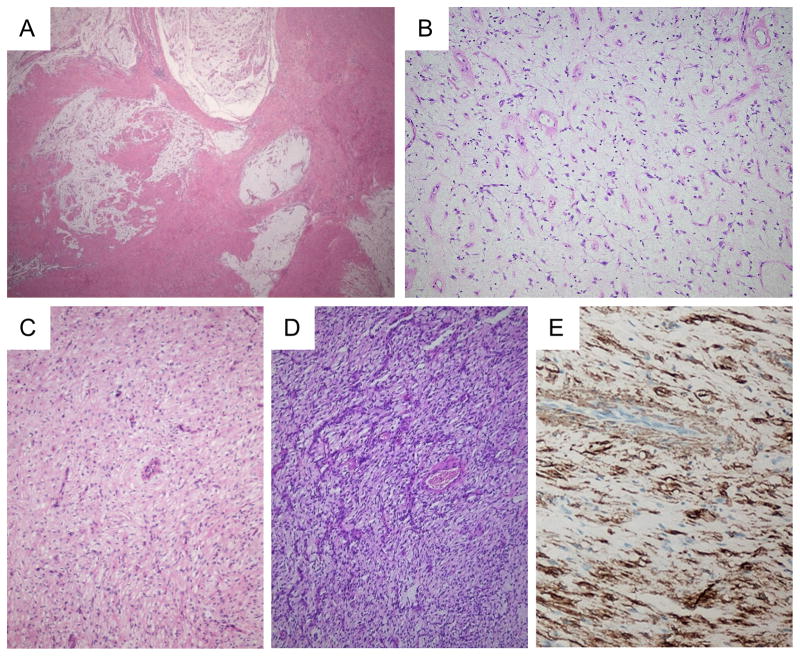
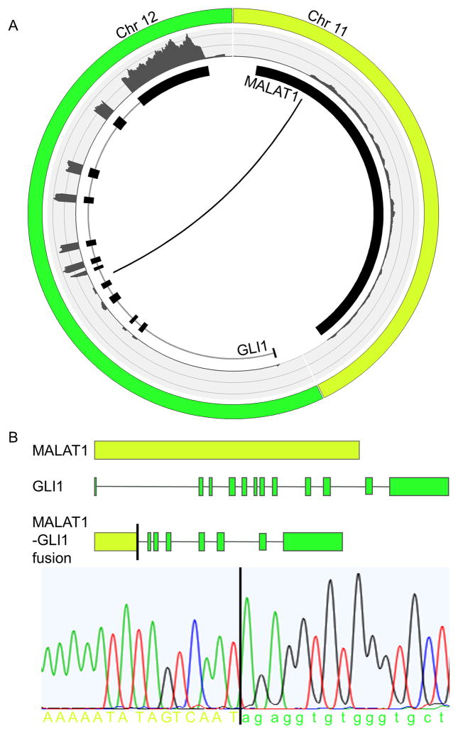
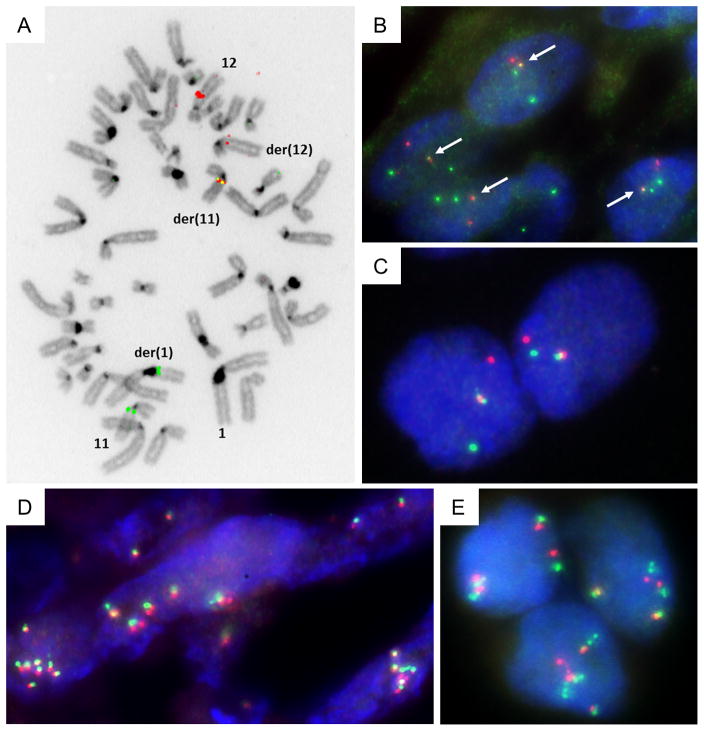
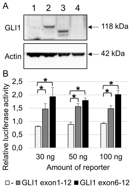
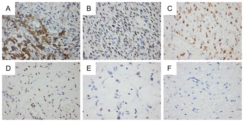
Plexiform fibromyxomas are rare neoplasms, being officially recognized as a distinct entity among benign mesenchymal gastric tumours in the 2010 WHO Classification of Tumours of the Digestive System. Characteristically, these tumours have a multinodular/plexiform growth pattern, and histologically contain variably cellular areas of bland myofibroblastic-type spindle cells embedded in an abundant myxoid matrix, rich in capillary-type vessels. As yet, the molecular and/or genetic features of these tumours are unknown. Here we describe a recurrent translocation, t(11;12)(q11;q13), involving the long non-coding gene metastasis-associated lung adenocarcinoma transcript 1 (MALAT1) and the gene glioma-associated oncogene homologue 1 (GLI1) in a subgroup of these tumours. The presence of the fusion transcript in our index case was confirmed using polymerase chain reaction (PCR) on genomic DNA, followed by Sanger sequencing. We showed that the truncated GLI1 protein is overexpressed and retains its capacity to transcriptionally activate its target genes. A specific FISH assay was developed to detect the novel MALAT1-GLI1 translocation in formalin-fixed, paraffin-embedded (FFPE) material. This resulted in the identification of two additional cases with this fusion and two cases with polysomy of the GLI1 gene. Finally, immunohistochemistry revealed that the GLI1 protein is exclusively overexpressed in those cases that harbour GLI1/12q13 genomic alterations. In conclusion, overexpression of GLI1 through a recurrent MALAT1-GLI1 translocation or GLI1 up-regulation delineates a pathogenically distinct subgroup of plexiform fibromyxomas with activation of the Sonic Hedgehog signalling pathway. Copyright © 2016 Pathological Society of Great Britain and Ireland. Published by John Wiley & Sons, Ltd.

Keywords: GLI1; MALAT1; oncogenic fusion; plexiform fibromyxoma.


Conflict of interest statement

Conflict of interest: The authors declare no conflicts of interest.

Figures

Figure 1
Figure 1
Histology of plexiform fibromyxomas of the stomach. Low power view, showing the lobular and plexiform architecture (A). At high power, the myxoid matrix contains numerous vessels, bland spindle cells and scattered inflammatory cells (B). In some lesions/nodules, the cellularity is somewhat increased. Note the bland appearance of the spindle cells and the prominent vessels (C and D). Tumor cells express alpha smooth muscle actin by IHC (E).
Figure 2
Figure 2
Graphical representation of the MALAT1-GLI1 fusion. The Circos plot depicts the interchromosomal translocation and its position relative to the different exons of GLI1 and the non-coding exon of MALAT1. The outer ring shows the FPKM counts representing the expression level of the different exons (A). Schematic representation of the MALAT1-GLI1 gene fusion. Structures for the genes have their basis in the UCSC reference sequences. For validation, a fusion-specific PCR followed by Sanger sequencing was performed on genomic DNA isolated from the tumor of index case 1 (B).
Figure 3
Figure 3
Metaphase dual-color FISH on index case 1 using SG-labeled RP11-472D15 and SO-labeled RP11-181L23 BAC probes that cover respectively the MALAT1 and GLI1 genes, indicate the presence of the MALAT1-GLI1 fusion (A). Interphase dual-color FISH on FFPE tissue from case 1 using SG-labeled RP11-472D15 and SO-labeled RP11-181L23 BAC probes that cover the MALAT1 and GLI1 genes, respectively. Overlapping green and red signals indicate MALAT1-GLI1 fusion (B). Interphase dual-color FISH on case 14 using SG-labeled RP11-1077C21 (centromeric) and SO-labeled RP11-571M06 (telomeric) BAC probes that flank the GLI1 gene. Rearrangement of GLI1 is pointed out by split apart green and red signals (C). Interphase dual-color FISH using probes that flank GLI1 on FFPE tissue from cases 4 and 15, showing polysomy of GLI1 (D and E).
Figure 4
Figure 4
Western blot showing overexpression of GLI1 protein in case 1 with the MALAT1-GLI1 fusion (lane 3). Case 2 without the fusion does not show GLI1 overexpression (lane 4). As a control, 293T cells were either untransfected (lane 1) or transfected with a full length GLI1 plasmid (lane 2) (A). Transcriptional activity of truncated GLI1 was tested using a dual-luciferase assay. 293T cells were transiently transfected with a luciferase reporter construct containing 11 copies of a GLI1 binding site together with a construct containing either full length GLI1 or only exons 6 to 12 of GLI1. Cells were lysed 48 hours after transfection. Results are represented as means of relative luciferase activity of 3 independent experiments performed in triplicate. Error bars indicate SEM; *p<0.05 (B).
Figure 5
Figure 5
Immunostaining for GLI1 protein expression. As a positive control with known GLI1 overexpression, a pericytoma carrying t(7;12)(p22;q13) and ACTB-GLI1 fusion showed diffuse cytoplasmic and nuclear immunopositivity (A). Plexiform fibromyxomas with MALAT1-GLI1 fusion (case 1 and 14, respectively) exhibit higher cellularity, and diffuse immunopositivity for GLI1 (B and C). Plexiform fibromyxoma with GLI1 polysomy (case 15), showing immunopositivity for GLI1 in a vast majority of tumor cells (D). Tumors without evident GLI1 involvement demonstrate either insubstantial positivity (case 2) or lack immunopositivity for GLI1 (case 13) (E and F).

Similar articles

Cited by

References

    1. Takahashi Y, Shimizu S, Ishida T, et al. Plexiform angiomyxoid myofibroblastic tumor of the stomach. Am J Surg Pathol. 2007;31:724–728. - PubMed
    1. Miettinen M, Makhlouf HR, Sobin LH, et al. Plexiform fibromyxoma: a distinctive benign gastric antral neoplasm not to be confused with a myxoid GIST. Am J Surg Pathol. 2009;33:1624–1632. - PubMed
    1. Ikemura M, Maeda E, Hatao F, et al. Plexiform angiomyxoid myofibroblastic tumor (PAMT) of the stomach. A case report focusing on its characteristic growth pattern. Int J Clin Exp Pathol. 2014;7:685–689. - PMC - PubMed
    1. Takahashi Y, Suzuki M, Fukusato T. Plexiform angiomyxoid myofibroblastic tumor of the stomach. World J Gastroenterol. 2010;16:2835–2840. - PMC - PubMed
    1. Yoshida A, Klimstra DS, Antonescu CR. Plexiform angiomyxoid tumor of the stomach. Am J Surg Pathol. 2008;32:1910–1912. author reply 1912–1913. - PubMed

Publication types

MeSH terms

Substances