. 2016 Jul:94:45-56.
doi: 10.1016/j.biomaterials.2016.03.048. Epub 2016 Apr 11.

Glypican-1 nanoliposomes for potentiating growth factor activity in therapeutic angiogenesis

Affiliations

Glypican-1 nanoliposomes for potentiating growth factor activity in therapeutic angiogenesis

Anthony J Monteforte et al. Biomaterials. 2016 Jul.

Abstract

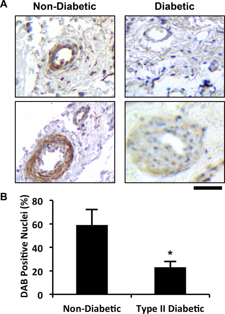
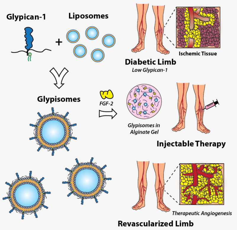
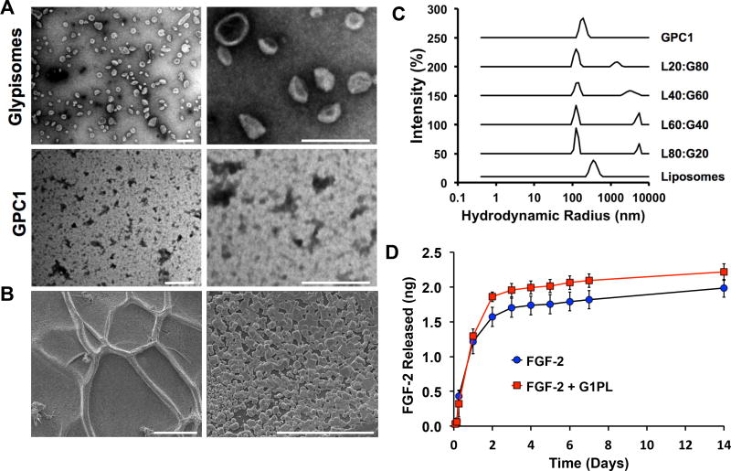
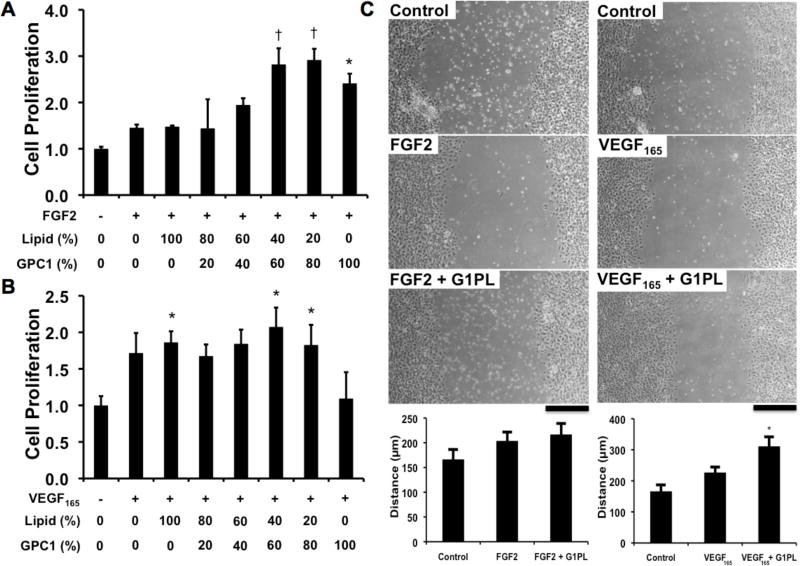
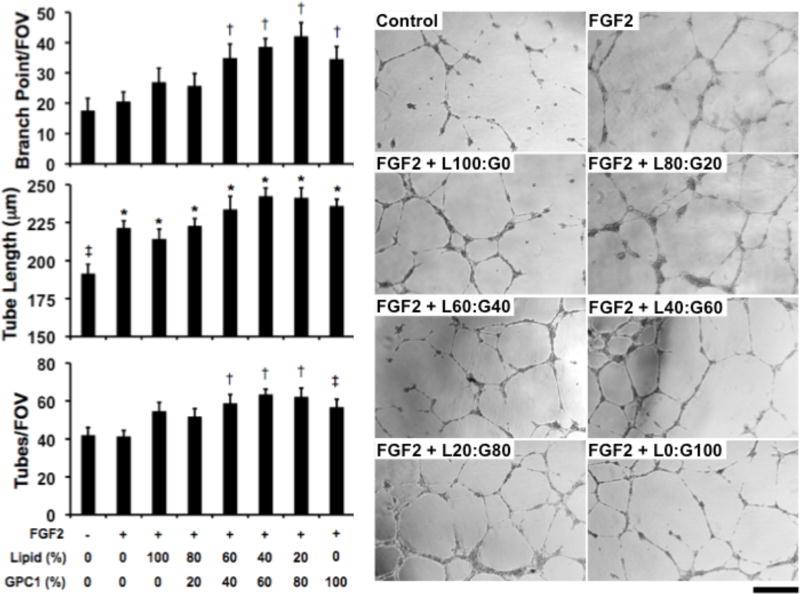
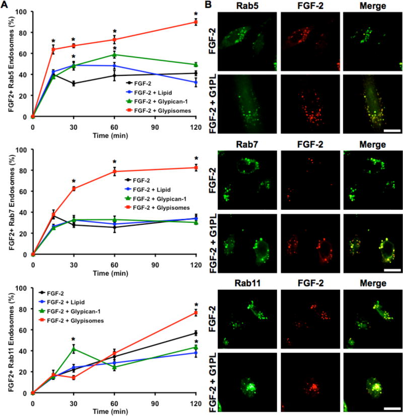
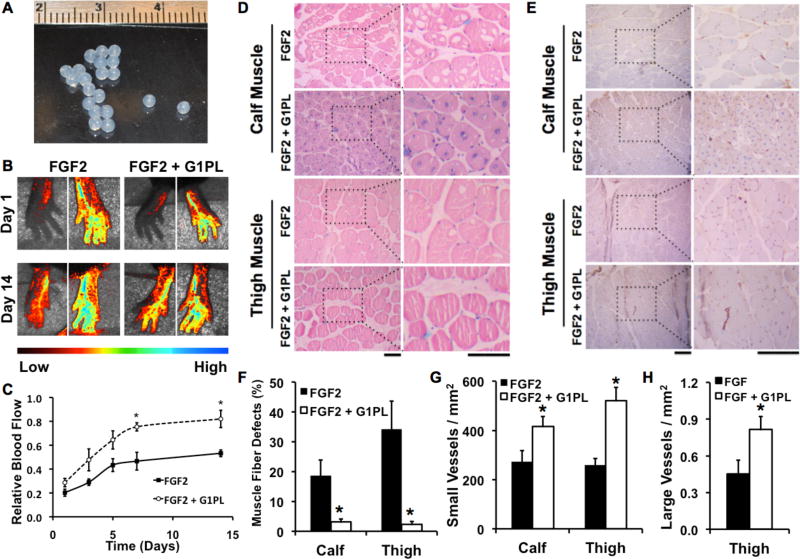
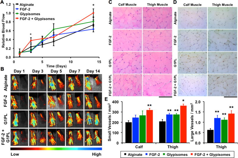
Therapeutic angiogenesis is a highly appealing concept for treating tissues that become ischemic due to vascular disease. A major barrier to the clinical translation of angiogenic therapies is that the patients that are in the greatest need of these treatments often have long term disease states and co-morbidities, such as diabetes and obesity, that make them resistant to angiogenic stimuli. In this study, we identified that human patients with type 2 diabetes have reduced levels of glypican-1 in the blood vessels of their skin. The lack of this key co-receptor in the tissue may make the application of exogenous angiogenic growth factors or cell therapies ineffective. We created a novel therapeutic enhancer for growth factor activity consisting of glypican-1 delivered in a nanoliposomal carrier (a "glypisome"). Here, we demonstrate that glypisomes enhance FGF-2 mediated endothelial cell proliferation, migration and tube formation. In addition, glypisomes enhance FGF-2 trafficking by increasing both uptake and endosomal processing. We encapsulated FGF-2 or FGF-2 with glypisomes in alginate beads and used these to deliver localized growth factor therapy in a murine hind limb ischemia model. Co-delivery of glypisomes with FGF-2 markedly increased the recovery of perfusion and vessel formation in ischemic hind limbs of wild type and diabetic mice in comparison to mice treated with FGF-2 alone. Together, our findings support that glypisomes are effective means for enhancing growth factor activity and may improve the response to local angiogenic growth factor therapies for ischemia.

Keywords: Angiogenesis; Fibroblast growth factor-2 (FGF-2); Glypican-1; Ischemia; Neovascularization; Peripheral arterial disease; Proteoliposomes; Vascular endothelial growth factor (VEGF).


Figures

Figure 1
Figure 1. Glypican-1 is reduced in mice and human with diabetes
(A) Immunostaining for glypican-1 in skin samples from diabetic and non-diabetic patients. (B) Analysis of the number of DAB positive nuclei within arterioles within the skin. *Statistically significant different from the no growth factor and growth factor alone groups (p < 0.05). Bar = 50 mm.
Figure 2
Figure 2
Overall diagram of glypisome-based therapeutics for the treatment of ischemia. Recombinant glypican-1 is embedded in a liposome membrane using detergent extraction. This construct is then combined with growth factors and polymerized into an alginate gel for delivery.
Figure 3
Figure 3. Characterization of glypisomes and alginate delivery gel
(A) Visualization of glypisomes and isolated glypican-1 (GPC1) by transmission electron microscopy. Bar = 200 nm. (B) Scanning electron microscopy visualization of the alginate gel after polymerization. Left bar = 100 µm and right bar = 30 µm. (C) Dynamic light scattering of isolated glypican-1 (GPC1), liposomes and glypisomes. Glypisomes are listed as composition ratio between lipid (L%) and protein (P%). (D) Release kinetics of FGF-2 from alginate gel with and without glypisomes added.
Figure 4
Figure 4. Glypisomes increase the proliferation and migration of endothelial cells
Proliferation of human endothelial cells after stimulation with growth factors and glypisomes with varying ratios between lipid and recombinant glypcian-1 (GPC-1). (A) Endothelial cells were treated with 10 ng/ml FGF-2 and glypisomes, and then proliferation was measured from Brdu incorposation assay. (B) Cells were treated with 10 ng/ml VEGF165 and glypicomes and then assayed for proliferation. *Statistically significant difference between group and the no growth factor group (p < 0.05). Statistically significant different from the no growth factor and growth factor alone groups (p < 0.05). (C) Wound closure after scratch wounding of an endothelial monolayer. Cells were treated with 10 ng/ml FGF-2 or 10 ng/ml VEGF at the time of injury. The glypisomes (G1PL) with a lipid to glypican-1 protein ratio of 20:80 were used. *Statistically significant different from the no growth factor and growth factor alone groups (p < 0.05). Bar = 200 µm.
Figure 5
Figure 5. Glypisomes enhance FGF-2 induced endothelial tube formation
Endothelial cells grown on growth factor reduced matrigel and the cells were treated with glypisomes with varying composition and 10 ng/ml FGF-2. After 16 hours the formation of tubes was assessed by phase contrast microscopy. *Statistically significant difference between group and the no growth factor group (p < 0.05). Statistically significant difference from the no growth factor and growth factor alone groups (p < 0.05). Bar = 200 µm.
Figure 6
Figure 6. Glypisomes increase endosomal trafficking of FGF-2 in HEK-293 cells
(A) Quantification of the percentage of Rab5, Rab7 or Rab11 positive (GFP) endosomes that contained FGF-2 after treatment of HEK-293 cells with various conditions. (B) Representative confocal images of cells treated with FGF-2 or FGF-2 with glypisomes at 120 min post treatment. *Statistically significant difference between the group and the FGF-2 only group at the same time point (p < 0.05). Bar = 20 µm.
Figure 7
Figure 7. Glypisomes enhance therapeutic angiogenesis with FGF-2 in hind limb ischemia
(A) Optimal composition glypisomes were encapsulated in alginate beads. (B) Ischemia was induced in the hind limb of mice by femoral artery ligation and alginate beads were implanted at the time of surgery. (C) Laser speckle contrast imaging was used to assess blood perfusion in the feet of the mice over time. Mice were given either alginate beads with FGF-2 or FGF-2 with glypisomes (G1PL). (D) Quantitative analysis of the perfusion of the feet after induction of hind limb ischemia. Relative blood flow was defined as the speckle contrast ratio between the ischemic limb and the control limb. *Statistically significant difference between group and FGF-2 alone group at the same time point (p < 0.05). (D) Histological analysis of the calf and thigh muscles of the ischemic limb after 14 days of treatment with FGF-2 or FGF-2 with glypisomes (G1PLs). (E) Immunohistochemical staining for blood vessels (PECAM-1) in the thigh and calf muscles. (F) Ischemic changes including the loss of muscle fibers was reduced in the calf muscle with FGF-2 and glypisome treatment in comparison to FGF-2 alone. Ischemic changes included the loss of muscle fibers/altered morphology with the tissue. (G) Quantification of small vessel density. (H) Quantification of large vessel density (diameter > 25 mm). *Statistically significant difference with FGF-2 alone group (p < 0.05). Bar = 100 µm.
Figure 8
Figure 8. Glypisomes enhance therapeutic angiogenesis when delivered with FGF-2 in an ob/ob hindlimb ischemia model
(A) Quantitative analysis of laser speckle contrast imaging gives relative blood flow analysis of perfusion measured over time. Relative flow defined as perfusion in the ischemic limb over the perfusion in the contralateral limb. (B) Representative laser speckle contrast images of each treatment group at 1, 3, 5, 7, and 14 days post injury. Right paw is from ischemic limb and left is the contralateral control. *Statistically significant difference with FGF-2 alone group (p < 0.05). (A) Representative H&E stained muscle fiber cross-sections of the calf and thigh of the ischemic limb. (B) Immunohistochemical staining for PECAM in calf and thigh muscle tissue of the ischemic limb. (C) Quantification of small vessel density in the calf and thigh muscle tissue, and large vessel density in the thigh muscle tissue. *Statistically significant difference from all other groups (p < 0.05). **Statistically significant difference from alginate group (p < 0.05). Bar = 100 µm.

References

    1. Norgren L, Hiatt WR, Dormandy JA, Nehler MR, Harris KA, Fowkes FG. Inter-Society Consensus for the Management of Peripheral Arterial Disease (TASC II) Journal of vascular surgery. 2007;(45 Suppl S):S5–67. - PubMed
    1. Udelson JE, Dilsizian V, Laham RJ, Chronos N, Vansant J, Blais M, et al. Therapeutic angiogenesis with recombinant fibroblast growth factor-2 improves stress and rest myocardial perfusion abnormalities in patients with severe symptomatic chronic coronary artery disease. Circulation. 2000;102:1605–10. - PubMed
    1. Lederman RJ, Mendelsohn FO, Anderson RD, Saucedo JF, Tenaglia AN, Hermiller JB, et al. Therapeutic angiogenesis with recombinant fibroblast growth factor-2 for intermittent claudication (the TRAFFIC study): a randomised trial. Lancet. 2002;359:2053–8. - PubMed
    1. Lederman RJ, Tenaglia AN, Anderson RD, Hermiller JB, Rocha-Singh K, Mendelsohn FO, et al. Design of the therapeutic angiogenesis with recombinant fibroblast growth factor-2 for intermittent claudication (TRAFFIC) trial. The American journal of cardiology. 2001;88:192–5. A6–7. - PubMed
    1. Rajagopalan S, Mohler ER, 3rd, Lederman RJ, Mendelsohn FO, Saucedo JF, Goldman CK, et al. Regional angiogenesis with vascular endothelial growth factor in peripheral arterial disease: a phase II randomized, double-blind, controlled study of adenoviral delivery of vascular endothelial growth factor 121 in patients with disabling intermittent claudication. Circulation. 2003;108:1933–8. - PubMed

MeSH terms