Leptin receptor null mice with reexpression of LepR in GnRHR expressing cells display elevated FSH levels but remain in a prepubertal state
- PMID: 27101301
- PMCID: PMC4935496
- DOI: 10.1152/ajpregu.00529.2015
Leptin receptor null mice with reexpression of LepR in GnRHR expressing cells display elevated FSH levels but remain in a prepubertal state
Abstract
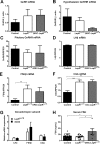
Leptin signals energy sufficiency to the reproductive hypothalamic-pituitary-gonadal (HPG) axis. Studies using genetic models have demonstrated that hypothalamic neurons are major players mediating these effects. Leptin receptor (LepR) is also expressed in the pituitary gland and in the gonads, but the physiological effects of leptin in these sites are still unclear. Female mice with selective deletion of LepR in a subset of gonadotropes show normal pubertal development but impaired fertility. Conditional deletion approaches, however, often result in redundancy or developmental adaptations, which may compromise the assessment of leptin's action in gonadotropes for pubertal maturation. To circumvent these issues, we adopted a complementary genetic approach and assessed if selective reexpression of LepR only in gonadotropes is sufficient to enable puberty and improve fertility of LepR null female mice. We initially assessed the colocalization of gonadotropin-releasing hormone receptor (GnRHR) and LepR in the HPG axis using GnRHR-IRES-Cre (GRIC) and LepR-Cre reporter (tdTomato or enhanced green fluorescent protein) mice. We found that GRIC and leptin-induced phosphorylation of STAT3 are expressed in distinct hypothalamic neurons. Whereas LepR-Cre was observed in theca cells, GRIC expression was rarely found in the ovarian parenchyma. In contrast, a subpopulation of gonadotropes expressed the LepR-Cre reporter gene (tdTomato). We then crossed the GRIC mice with the LepR null reactivable (LepR(loxTB)) mice. These mice showed an increase in FSH levels, but they remained in a prepubertal state. Together with previous findings, our data indicate that leptin-selective action in gonadotropes serves a role in adult reproductive physiology but is not sufficient to allow pubertal maturation in mice.
Keywords: HPG axis; gonadotropes; obesity; puberty; sexual maturation.
Copyright © 2016 the American Physiological Society.
Figures


Similar articles
-
Leptin Signaling in AgRP Neurons Modulates Puberty Onset and Adult Fertility in Mice.J Neurosci. 2017 Apr 5;37(14):3875-3886. doi: 10.1523/JNEUROSCI.3138-16.2017. Epub 2017 Mar 8. J Neurosci. 2017. PMID: 28275162 Free PMC article.
-
Selective deletion of leptin receptors in gonadotropes reveals activin and GnRH-binding sites as leptin targets in support of fertility.Endocrinology. 2014 Oct;155(10):4027-42. doi: 10.1210/en.2014-1132. Epub 2014 Jul 24. Endocrinology. 2014. PMID: 25057790 Free PMC article.
-
Leptin's effect on puberty in mice is relayed by the ventral premammillary nucleus and does not require signaling in Kiss1 neurons.J Clin Invest. 2011 Jan;121(1):355-68. doi: 10.1172/JCI45106. Epub 2010 Dec 22. J Clin Invest. 2011. PMID: 21183787 Free PMC article.
-
A critical view of the use of genetic tools to unveil neural circuits: the case of leptin action in reproduction.Am J Physiol Regul Integr Comp Physiol. 2014 Jan 1;306(1):R1-9. doi: 10.1152/ajpregu.00444.2013. Epub 2013 Nov 6. Am J Physiol Regul Integr Comp Physiol. 2014. PMID: 24196667 Free PMC article. Review.
-
Hypothalamic sites of leptin action linking metabolism and reproduction.Neuroendocrinology. 2011;93(1):9-18. doi: 10.1159/000322472. Epub 2010 Nov 24. Neuroendocrinology. 2011. PMID: 21099209 Free PMC article. Review.
Cited by
-
Sexually dimorphic distribution of Prokr2 neurons revealed by the Prokr2-Cre mouse model.Brain Struct Funct. 2017 Dec;222(9):4111-4129. doi: 10.1007/s00429-017-1456-5. Epub 2017 Jun 14. Brain Struct Funct. 2017. PMID: 28616754 Free PMC article.
-
Adipokines (Leptin, Adiponectin, Resistin) Differentially Regulate All Hormonal Cell Types in Primary Anterior Pituitary Cell Cultures from Two Primate Species.Sci Rep. 2017 Mar 6;7:43537. doi: 10.1038/srep43537. Sci Rep. 2017. PMID: 28349931 Free PMC article.
-
Lack of AR in LepRb Cells Disrupts Ambulatory Activity and Neuroendocrine Axes in a Sex-Specific Manner in Mice.Endocrinology. 2020 Aug 1;161(8):bqaa110. doi: 10.1210/endocr/bqaa110. Endocrinology. 2020. PMID: 32609838 Free PMC article.
-
Gonadotrope-specific deletion of the BMP type 2 receptor does not affect reproductive physiology in mice†‡.Biol Reprod. 2020 Mar 13;102(3):639-646. doi: 10.1093/biolre/ioz206. Biol Reprod. 2020. PMID: 31724029 Free PMC article.
-
The Importance of Leptin to Reproduction.Endocrinology. 2021 Feb 1;162(2):bqaa204. doi: 10.1210/endocr/bqaa204. Endocrinology. 2021. PMID: 33165520 Free PMC article. Review.
References
-
- Barash IA, Cheung CC, Weigle DS, Ren H, Kabigting EB, Kuijper JL, Clifton DK, Steiner RA. Leptin is a metabolic signal to the reproductive system. Endocrinology 137: 3144–3147, 1996. - PubMed
-
- Batt RAL, Everard DM, Gillies G, Wilkinson M, Wilson CA, Yeo TA. Investigation into the hypogonadism of the obese mouse (genotype ob/ob). J Reprod Fertil 64: 363–371, 1982. - PubMed
Publication types
MeSH terms
Substances
Grants and funding
LinkOut - more resources
Full Text Sources
Other Literature Sources
Medical
Molecular Biology Databases
Miscellaneous

