. 2016 Jun 1;9(6):607-20.
doi: 10.1242/dmm.024661. Epub 2016 Apr 21.

Musculocontractural Ehlers-Danlos syndrome and neurocristopathies: dermatan sulfate is required for Xenopus neural crest cells to migrate and adhere to fibronectin

Affiliations

Musculocontractural Ehlers-Danlos syndrome and neurocristopathies: dermatan sulfate is required for Xenopus neural crest cells to migrate and adhere to fibronectin

Nadège Gouignard et al. Dis Model Mech. .

Abstract

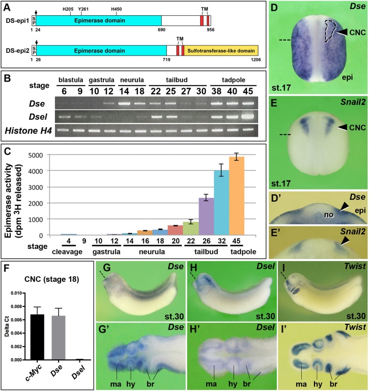
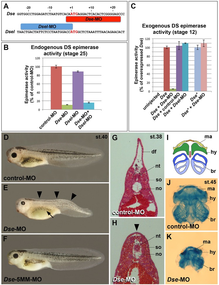
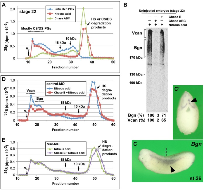
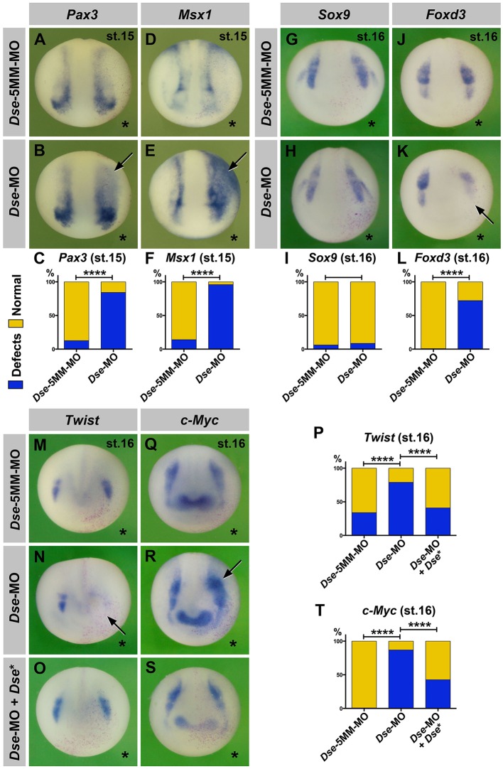
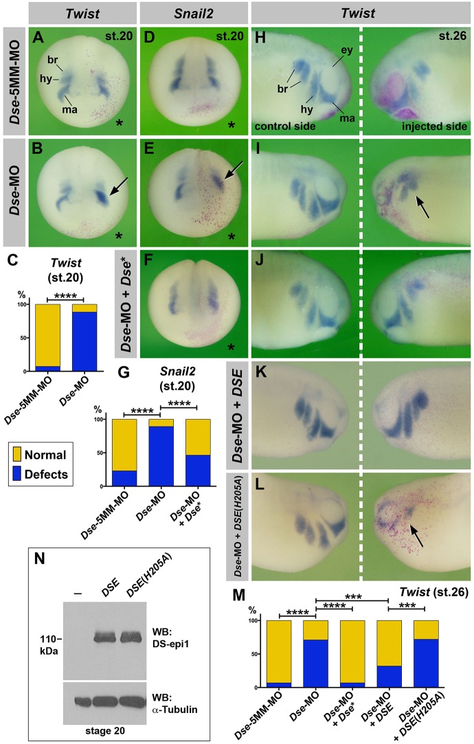
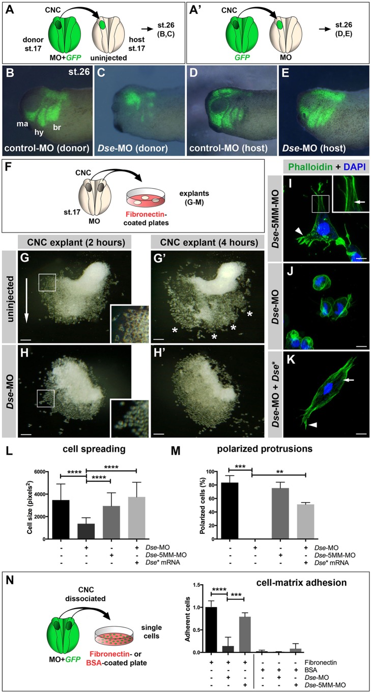
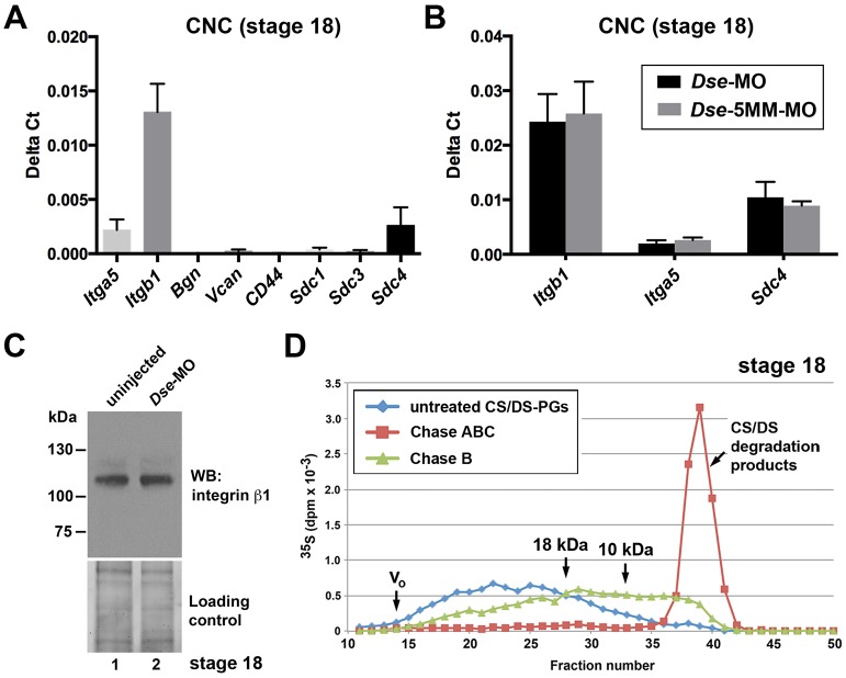
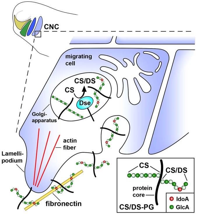
Of all live births with congenital anomalies, approximately one-third exhibit deformities of the head and face. Most craniofacial disorders are associated with defects in a migratory stem and progenitor cell population, which is designated the neural crest (NC). Musculocontractural Ehlers-Danlos syndrome (MCEDS) is a heritable connective tissue disorder with distinct craniofacial features; this syndrome comprises multiple congenital malformations that are caused by dysfunction of dermatan sulfate (DS) biosynthetic enzymes, including DS epimerase-1 (DS-epi1; also known as DSE). Studies in mice have extended our understanding of DS-epi1 in connective tissue maintenance; however, its role in fetal development is not understood. We demonstrate that DS-epi1 is important for the generation of isolated iduronic acid residues in chondroitin sulfate (CS)/DS proteoglycans in early Xenopus embryos. The knockdown of DS-epi1 does not affect the formation of early NC progenitors; however, it impairs the correct activation of transcription factors involved in the epithelial-mesenchymal transition (EMT) and reduces the extent of NC cell migration, which leads to a decrease in NC-derived craniofacial skeleton, melanocytes and dorsal fin structures. Transplantation experiments demonstrate a tissue-autonomous role for DS-epi1 in cranial NC cell migration in vivo Cranial NC explant and single-cell cultures indicate a requirement of DS-epi1 in cell adhesion, spreading and extension of polarized cell processes on fibronectin. Thus, our work indicates a functional link between DS and NC cell migration. We conclude that NC defects in the EMT and cell migration might account for the craniofacial anomalies and other congenital malformations in MCEDS, which might facilitate the diagnosis and development of therapies for this distressing condition. Moreover, the presented correlations between human DS-epi1 expression and gene sets of mesenchymal character, invasion and metastasis in neuroblastoma and malignant melanoma suggest an association between DS and NC-derived cancers.

Keywords: Cancer; Cell migration; Dermatan sulfate; Musculocontractural Ehlers–Danlos syndrome; Neural crest; Xenopus.


Conflict of interest statement

Competing interests

The authors declare no competing or financial interests.

Figures

Fig. 1.
Fig. 1.
Expression and activity of the two dermatan sulfate epimerases in Xenopus embryos. (A) Protein structures. Xenopus DS-epi1 and DS-epi2 contain cleavable signal peptides (SP, arrows), an epimerase domain and two transmembrane (TM) domains. In DS-epi1, the catalytic residues His205, Tyr261 and His450 are indicated, which are also conserved in DS-epi2. DS-epi2 contains an additional sulfotransferase-like domain. (B) RT-PCR analysis of Dse and Dsel. Histone H4 is used as the loading control. A minimum of two experiments (n≥2) was performed with three independent biological samples. (C) DS epimerase activity in early Xenopus embryos. Results are mean±s.d. from triplicates (two independent experiments). (D-E′) Whole-mount in situ hybridization of neurula embryos in the dorsal view (D,E) and in transversal section (D′,E′). The arrowheads label the pre-migratory CNC cells. The region enclosed by the dashed line demarcates the Snail2+ CNC embedded in the Dse expression domain. (F) qPCR analysis in CNC explants at stage 18. c-Myc was used as a CNC cell marker. Note that Dse but not Dsel mRNA is detected. Results are mean±s.d. from triplicates (n=4 biological replicates). (G-I′) Tailbud embryos in the lateral view (G-I) and horizontal section (G′-I′). Note that Dse and Dsel overlap with Twist expression in migrating CNC cells. Section planes are indicated with dashed straight lines. br, branchial arch segments; epi, epidermis; hy, hyoid segment; ma, mandibular segment; no, notochord.
Fig. 2.
Fig. 2.
Knockdown of DS-epi1 reduces DS epimerase activity and NC-derived structures. (A) Morpholino oligonucleotides target the translation initiation sites of Dse and Dsel. (B) Endogenous DS epimerase activity is substantially decreased by Dse-MO but only a little by Dsel-MO in stage 25 embryos. (C) Epimerase activity induced by the injection of 1 ng Dse mRNA is blocked by Dse-MO, but not by control-MO and Dsel-MO. The activity of 1 ng non-targeted Dse* mRNA is not affected by Dse-MO. Results in B and C are mean±s.d. (n=3). (D) Tadpole at stage 40 injected with control-MO. (E,F) Microinjection of Dse-MO, but not Dse-5MM-MO, induces small eyes, a lack of dorsal fin structures (arrowheads) and reduced melanocyte formation (arrow). (G,H) Transversal trunk sections of stage 38 embryos following hematoxylin and eosin staining. Note the lack of a dorsal fin (arrowhead), dorsally approaching somites and hypoplastic notochord in the Dse-morphant embryo. (I-K) Ventral view of head skeletons at stage 45 in a schematic overview (I) and following Alcian Blue staining (J,K). Injection of Dse-MO, but not control-MO, causes a reduction of NC-derived cartilage structures. br, branchial segment; hy, hyoid segment; df, dorsal fin; ma, mandibular segment; no, notochord; nt, neural tube; so, somite. The proportion of examined tadpoles or explants with the indicated phenotype was as follows: D, 70/70; E, 71/114 (microcephaly), 92/114 (reduced dorsal fin), 70/114 (less melanocytes); F, 63/63; G, 4/4; H, 4/4; J, 25/25; and K, 20/20.
Fig. 3.
Fig. 3.
Presence of IdoA in CS/DS PGs of early embryos. (A) At stage 22, IdoA is present in high molecular mass CS/DS PGs, as demonstrated by the size-fractionation of [35S]-containing PGs. The HS and CS/DS degradation products are produced by nitrous acid and Chase ABC treatment, respectively. CS/DS PGs represent 72% of the high molecular mass PGs (fractions 12-17). (B) SDS-PAGE analysis of [35S]-labeled CS/DS PGs. The same samples analyzed by gel filtration in A were separated using a 4-10% gradient SDS-PAGE following nitrous acid or, alternatively, Chase ABC or Chase B treatments. The fluography indicates CS/DS PGs (brackets) with an apparent molecular mass of 200-300 kDa (Bgn) and ∼1000 kDa (Vcan). The percentages of radioactivity in the framed areas are indicated below each lane. (C,C′) Whole-mount in situ hybridization of Bgn at stage 26. Embryo is shown in the lateral view (C) and transversally sectioned (C′). Arrowheads indicate migrating trunk neural crest cells. The section planes are indicated by the dashed straight line. (D,E) Chase B treatment degrades CS/DS chains in high molecular mass PGs in control-MO-injected embryos (D) but not Dse-MO-injected embryos (E). Bgn, biglycan; Chase, chondroitinase; Vcan, versican.
Fig. 4.
Fig. 4.
DS-epi1 regulates gene markers of the neural plate border and CNC. Whole-mount in situ hybridization of early neurula embryos in an anterior view. The injected side is marked with a star. (A-F) A single injection of Dse-MO into embryos causes expansion of Pax3 and Msx1 expression at the neural plate border (arrows). The Dse-5MM-MO has no effect. A quantification of the percentage of embryos with defects is shown in C and F. (G-T) Dse-MO has no significant effect on Sox9; however, it triggers a reduction in Foxd3 and Twist expression, as well as an expansion of c-Myc expression (arrows). Normal Twist and c-Myc expression is restored by the co-injection of Dse-MO and 250 pg Dse* mRNA. nlacZ mRNA was injected as a lineage tracer (red nuclei). A quantification of the percentage of embryos with defects is shown in I, L, P and T. The proportion of examined embryos with the indicated phenotype was as follows: A, 37/42; B, 32/38; D, 26/30; E, 30/31; G, 35/37; H, 58/63; J, 30/30; K, 26/36; M, 13/20; N, 48/61; O, 16/27; Q, 90/90; R, 77/89; and S, 15/26. ****P<0.0001 (Fisher's exact test with two-tailed P-value calculation).
Fig. 5.
Fig. 5.
DS-epi1 regulates CNC cell migration. (A-G) Anterior view of late neurula embryos. The injected side is marked with a star. Dse-MO impairs the segregation of Twist+ and Snail2+ CNC cells (arrows). The effect is reversed by 250 pg Dse* mRNA. A quantification of the percentage of embryos with defects is shown in C and G. (H-M) Lateral view of tailbud embryos. Dse-MO leads to defective migration of Twist+ CNC cells (arrow) on the injected side, which is rescued by the co-injection of 250 pg Dse* mRNA and 25 pg pcDNA3/CTAP-DSE plasmid, but not 25 pg pcDNA3/CTAP-DSE (H205A) plasmid DNA. A quantification of the percentage of embryos with defects is shown in M. (N) Western blot analysis of lysates from embryos injected with 100 pg pcDNA3/CTAP-DSE or pcDNA3/CTAP-DSE (H205A) plasmid DNA and probed for DS-epi1. α-tubulin is a loading control. br, branchial segment; ey, eye; hy, hyoid segment; ma, mandibular segment. The proportion of examined embryos with the indicated phenotype was as follows: A, 15/16; B, 30/34; D, 41/46; E, 50/65; F, 20/37; H, 25/27; I, 31/41; J, 36/44; K, 27/40; and L, 14/18. ***P<0.005; ****P<0.0001 (Fisher's exact test with two-tailed P-value calculation).
Fig. 6.
Fig. 6.
DS-epi1 has a tissue-autonomous role in CNC cell migration, adherence to fibronectin and cell polarization. (A,A′) Schemes for transplantation experiments. A CNC explant from an embryo injected with 300 pg GFP mRNA was homotypically grafted at stage 17. MOs were injected into the donor (A) or host embryo (A′). (B-E) Lateral view of embryos at stage 26. Grafted GFP+ CNC cells migrate ventrally when derived from control-MO-injected embryos (B); however, they do not properly migrate when derived from Dse-MO-injected embryos (C). br, branchial segment; hy, hyoid segment; ma, mandibular segment. The CNC cell migration was normal when the host embryo was injected with control-MO or Dse-MO (D,E). Three independent experiments were performed (n=3). (F) Scheme illustrating the culture of stage 17 morphant CNC explants on fibronectin-coated plates. (G,G′) At 2 h after plating (G), the control-MO-injected CNC explant exhibits collective cell migration in one direction (arrow). The inset shows a magnification of spread cells. After 4 h (G′), the cells migrate in distinct streams (asterisks). (H,H′) Cells of Dse-MO-injected CNC explants detach from each other and fail to adhere to the fibronectin substrate. The inset depicts a magnification of the spherical cells. (I-K) Confocal microscopy of fixed CNC cells after 5 h of explant culture on fibronectin. Phalloidin–Alexa-Fluor-488 and DAPI label F-actin and cell nuclei, respectively. The Dse-5MM-MO-injected control cell (I) exhibits lamellipodia at the leading edge (arrowhead) and stress fibers in the inner regions of the cell (arrow in inset). Dse-morphant cells (J) exhibit cortical networks of stress fibers and lack polarized protrusions. Co-injection of Dse-MO and 1 ng Dse* mRNA per embryo (K) restores the normal cytoskeleton and cell shape. (L,M) Quantification of cell spreading (L) and formation of polarized cell protrusions (M) in dissociated phalloidin-stained single cells from CNC explants following 5 h of culture on fibronectin. Cell spreading and polarized protrusions were quantified by calculating the cell size as the square number of pixels (ImageJ) and determining the percentage of cells with lamellipodia or filopodia, respectively. Uninjected and Dse-5MM-MO-injected explants exhibit a similar extent of cell spreading and formation of polarized protrusions. The reduction in the cell size and the lack of lamellipodia and filopodia are rescued by the co-injection of Dse* mRNA in Dse-morphant explants. A minimum of 100 cells per sample were evaluated in each experiment. Number of independent experiments (n≥3). Results are mean±s.d. (N) Cell–matrix adhesion of dissociated single CNC cells on fibronectin- or BSA-coated plates. Following the co-injection of MO and 300 pg GFP mRNA, CNC explants from stage 17 embryos were dissociated in Ca2+- and Mg2+-free medium and cultured for 45 min on fibronectin or BSA. The Dse-morphant cells exhibit decreased adhesion to fibronectin compared with the control and Dse-5MM-MO-injected cells. None of the analyzed cell samples exhibited significant cell adhesion to BSA. At least three independent experiments were performed for each sample (n≥3). Results are mean±s.d. The proportion of examined explants or cells with the indicated phenotype was as follows: B, 10/12; C, 11/13; D, 7/7; E, 9/9; G, 30/34; H, 26/28. Scale bars: 100 µm (G-H′); 10 µm (I-K). **P<0.01, ***P<0.001, ****P<0.0001 (one-way ANOVA multiple comparisons test with Tukey correction).
Fig. 7.
Fig. 7.
CS/DS-PGs in CNC cells. (A) qPCR analysis in uninjected CNC explants at stage 18. Note abundant expression of Itga5, Itgb1 and Sdc4. Results are mean±s.d. from triplicates (n≥4 biological replicates). (B) Dse-MO does not differentially affect the mRNA levels of Itga5, Itgb1 and Sdc4 compared with Dse-5MM-MO. Results are mean±s.d. (n≥4 biological replicates). (C) Dse-MO does not reduce the protein amount of integrin β1 in explants enriched in neural crest and epidermis of stage 18 embryos. Western blotting was performed on a 7.5% Mini-Protean TGX Stain-free gel (Bio-Rad). The loading control was ascertained prior to blotting using the ChemiDoc Touch Imaging System. Resuts is representative of two independent experiments (n=2). (D) Metabolic labeling of PGs in stage 18 CNC explants. Note that Chase B partially degrades CS/DS PGs >18 kDa. The IdoA is a rare modification because the split chains are ∼10 kDa.
Fig. 8.
Fig. 8.
Model for the stimulation of CNC cell migration by CS/DS PGs in a post-neurula embryo. DS-epi1 converts GlcA into isolated IdoA residues on CS/DS PGs. The interaction between CS/DS PGs and extracellular fibronectin stimulates cytoskeletal rearrangement and polarized cell migration.

References

    1. Alfandari D., Cousin H., Gaultier A., Hoffstrom B. G. and DeSimone D. W. (2003). Integrin α5β1 supports the migration of Xenopus cranial neural crest on fibronectin. Dev. Biol. 260, 449-464. 10.1016/S0012-1606(03)00277-X - DOI - PubMed
    1. Bao X., Muramatsu T. and Sugahara K. (2005). Demonstration of the pleiotrophin-binding oligosaccharide sequences isolated from chondroitin sulfate/dermatan sulfate hybrid chains of embryonic pig brains. J. Biol. Chem. 280, 35318-35328. 10.1074/jbc.M507304200 - DOI - PubMed
    1. Barriga E. H., Maxwell P. H., Reyes A. E. and Mayor R. (2013). The hypoxia factor Hif-1α controls neural crest chemotaxis and epithelial to mesenchymal transition. J. Cell Biol. 201, 759-776. 10.1083/jcb.201212100 - DOI - PMC - PubMed
    1. Bartolini B., Thelin M. A., Rauch U., Feinstein R., Oldberg Å., Malmström A. and Maccarana M. (2012). Mouse development is not obviously affected by the absence of dermatan sulfate epimerase 2 in spite of a modified brain dermatan sulfate composition. Glycobiology 22, 1007-1016. 10.1093/glycob/cws065 - DOI - PubMed
    1. Bartolini B., Thelin M. A., Svensson L., Ghiselli G., van Kuppevelt T. H., Malmström A. and Maccarana M. (2013). Iduronic acid in chondroitin/dermatan sulfate affects directional migration of aortic smooth muscle cells. PLoS ONE 8, e66704 10.1371/journal.pone.0066704 - DOI - PMC - PubMed

Publication types

MeSH terms