. 2016 Apr 22:6:24805.
doi: 10.1038/srep24805.

Bone marrow-derived mesenchymal stem cells improve diabetes-induced cognitive impairment by exosome transfer into damaged neurons and astrocytes

Affiliations

Bone marrow-derived mesenchymal stem cells improve diabetes-induced cognitive impairment by exosome transfer into damaged neurons and astrocytes

Masako Nakano et al. Sci Rep. .

Abstract

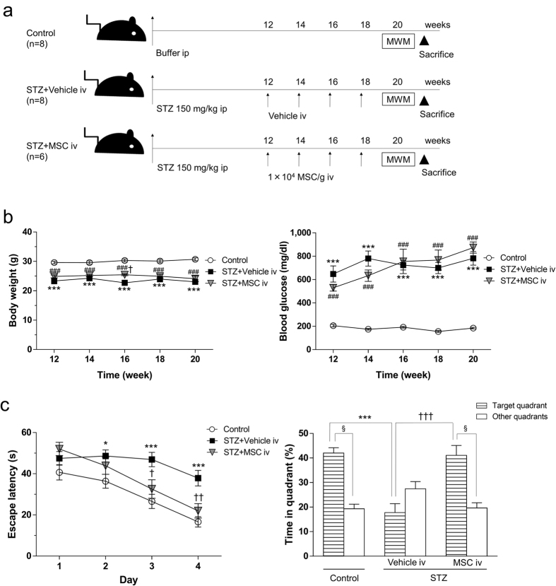
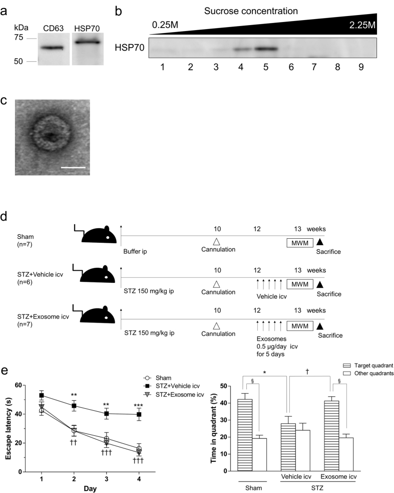
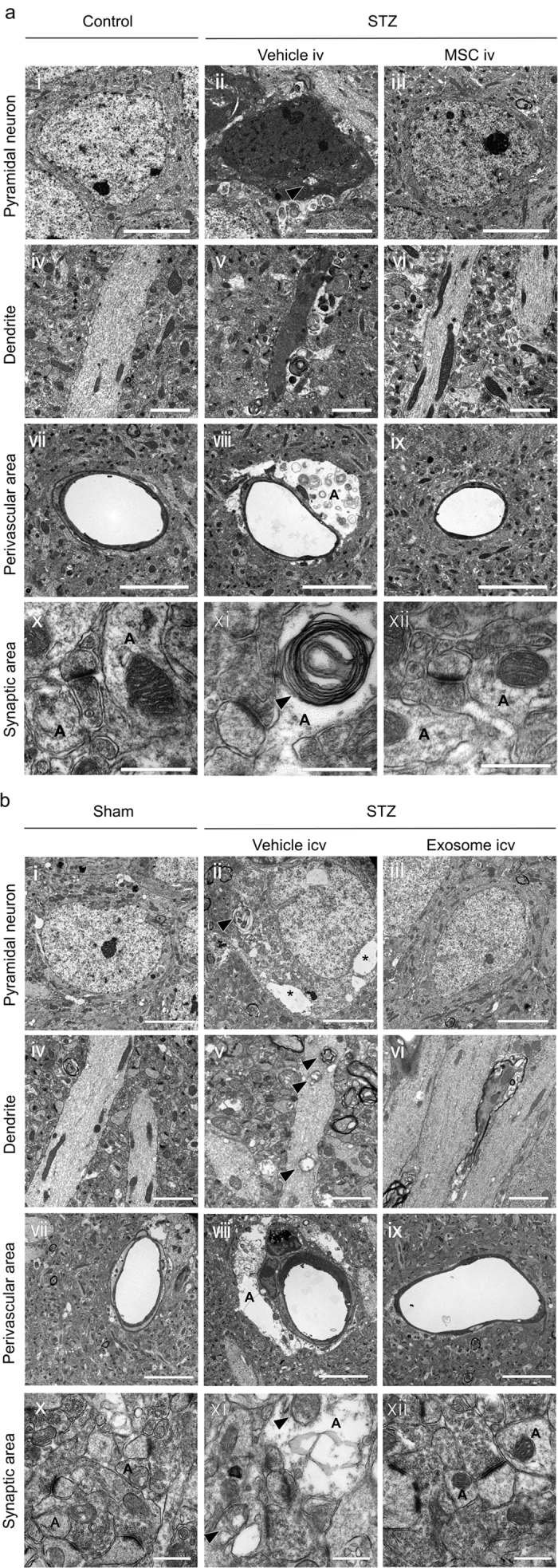
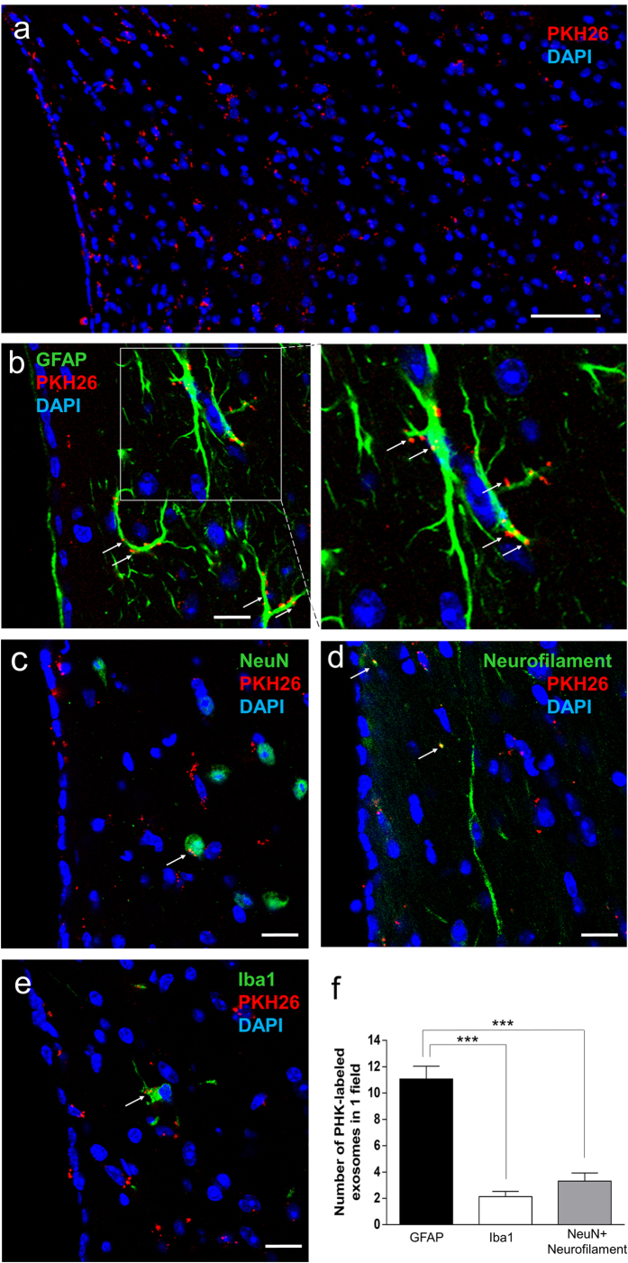
The incidence of dementia is higher in diabetic patients, but no effective treatment has been developed. This study showed that rat bone marrow mesenchymal stem cells (BM-MSCs) can improve the cognitive impairments of STZ-diabetic mice by repairing damaged neurons and astrocytes. The Morris water maze test demonstrated that cognitive impairments induced by diabetes were significantly improved by intravenous injection of BM-MSCs. In the CA1 region of the hippocampus, degeneration of neurons and astrocytes, as well as synaptic loss, were prominent in diabetes, and BM-MSC treatment successfully normalized them. Since a limited number of donor BM-MSCs was observed in the brain parenchyma, we hypothesized that humoral factors, especially exosomes released from BM-MSCs, act on damaged neurons and astrocytes. To investigate the effectiveness of exosomes for treatment of diabetes-induced cognitive impairment, exosomes were purified from the culture media and injected intracerebroventricularly into diabetic mice. Recovery of cognitive impairment and histological abnormalities similar to that seen with BM-MSC injection was found following exosome treatment. Use of fluorescence-labeled exosomes demonstrated that injected exosomes were internalized into astrocytes and neurons; these subsequently reversed the dysfunction. The present results indicate that exosomes derived from BM-MSCs might be a promising therapeutic tool for diabetes-induced cognitive impairment.


Figures

Figure 1
Figure 1. Intravenous injection of BM-MSCs into STZ-induced diabetic mice.
(a) Experimental protocol. At 12 weeks after STZ injection, mice are injected iv with 1 × 104 MSCs/g body weight four times every 2 weeks or with PBS for vehicle injection. Two weeks after the last injection, the Morris water maze (MWM) test is carried out. (b) Changes in body weight and serum blood glucose levels after MSC injection. ***P < 0.001, STZ + vehicle vs. control; ###P < 0.001, STZ + MSC vs. control; P < 0.05, STZ + vehicle vs. STZ + MSC, two-way ANOVA, Bonferroni post-test. Values are means ± s.e.m, n = 6–8. (c) MWM test. In the hidden training test, STZ + vehicle mice exhibit longer escape latency on days 2, 3 and 4 than control mice. The escape latency of STZ + MSC mice is shortened on days 3 and 4 compared to STZ + vehicle mice. *P < 0.05, ***P < 0.001, STZ + vehicle vs. control; P < 0.05, ††P < 0.01, STZ + vehicle vs. STZ + MSC, two-way ANOVA (F(2, 85) = 16.38 P < 0.0001). Values are means ± s.e.m, n = 6-8. In the probe test, the target quadrant occupancy of STZ + vehicle mice is significantly reduced compared with control mice (***P < 0.001, one-way ANOVA, Bonferroni post-test). The target quadrant occupancy of STZ + MSC mice is significantly elevated compared to STZ + vehicle mice. (†††P < 0.001, one-way ANOVA, Bonferroni post-test). Control and STZ + MSC mice spend significantly more time in the target quadrant than any of the other quadrants (§P < 0.0001, unpaired two-tailed t-test). For STZ + vehicle mice, the time spent in the target quadrant is similar to that spent in the other quadrants (P = 0.1406, unpaired two-tailed t-test).
Figure 2
Figure 2. Isolation of BM-MSC-derived exosomes and intracerebroventricular injection of BM-MSC-derived exosomes into STZ-induced diabetic mice.
(a) Western blot analysis for pellets obtained after 100,000 × g centrifugation. Positive reactions for CD63 and HSP70 are detected. (b) Further purification by a sucrose step gradient. HSP70 is detected at ranges between 1.11–1.15 g/mL (fractions 4–5). (c) Electron microscopic observation of an exosome. Bar, 50 nm. (d) Experimental protocols. At 12 weeks after STZ injection, mice are injected icv with 0.5 μg of exosomes in 2 μL aCSF once a day for 5 successive days, or with 2 μL aCSF for vehicle injection. Two days after the last injection, MWM tests are carried out. (e) MWM test. In the hidden training test, STZ + vehicle mice exhibit longer escape latency on days 2, 3 and 4 than sham-operated mice. The escape latency of STZ + exosome mice is shortened on days 2, 3 and 4 compared to STZ + vehicle mice. **P < 0.01, ***P < 0.001 STZ + vehicle vs. sham; ††P < 0.01, †††P < 0.001, STZ + vehicle vs. STZ + exosome; two-way ANOVA (F(2,77) = 23.65, P < 0.0001). Values are means ± s.e.m, n = 6–7. In the probe test, the target quadrant occupancy of STZ + vehicle mice is significantly reduced (*P < 0.05, one-way ANOVA, Bonferroni post-test). The target quadrant occupancy of STZ + exosome mice is significantly higher than of STZ + vehicle mice. (P < 0.05, one-way ANOVA, Bonferroni post-test). Sham and STZ + exosome mice spend significantly more time in the target quadrant than any of the other quadrants (§P < 0.0001, unpaired two-tailed t-test). For STZ + vehicle mice, the time spent in the target quadrant is similar to that spent in the other quadrants (P = 0.1406, unpaired two-tailed t-test).
Figure 3
Figure 3. Morphological analysis in the CA1 region.
(a,f) The number of NeuN-positive cells in STZ + vehicle mice is significantly decreased compared to control mice, and the number is not recovered in STZ + MSC and STZ + exosome mice. Bar, 50 μm. (b,g) The staining intensity of 4HNE(4-hydroxynonenal), an oxidative stress marker, is significantly increased in the dendrites (labeled with MAP2) of CA1 neurons in STZ + vehicle mice, and the increase is recovered to normal levels in STZ + MSC and STZ + exosome mice. There is no difference in the MAP2 area among the three groups. Bar, 50 μm. (c,h) The density of synaptophysin is significantly decreased in STZ + vehicle mice, and this decrease is recovered in STZ + MSC and STZ + exosome mice. Bar, 50 μm. (d,i) The number of Iba1-positive microglia is significantly increased in the STZ + vehicle group, and this increase recovers to normal levels in STZ + MSC and STZ + exosome mice. Bar, 100 μm. (e,j) The number of GFAP-positive astrocytes is not different among the three groups, but the area of the GFAP-positive reaction is decreased in STZ + vehicle mice. This decrease is not recovered in STZ + MSC mice nor in STZ + exosome mice. Bar, 100 μm. (a–j) *P < 0.05, **P < 0.01, ***P < 0.001, one-way ANOVA, Bonferroni post-test. Values are means ± s.e.m, n = 3–4.
Figure 4
Figure 4. Electron microscopic study.
(a i–iii) In the pyramidal layer, dark neurons with swollen mitochondria (arrowhead) are prominent in STZ + vehicle mice. These abnormalities have completely disappeared in STZ + MSC mice. Bar, 5 μm. (b i–iii) In the cytoplasm of pyramidal neurons, swollen mitochondria and vacuolization (*) are prominent in STZ + vehicle mice. These abnormalities have completely disappeared in STZ + exosome mice. Bar, 5 μm. (a iv–vi) While dark and shrunk dendrites are seen in STZ + vehicle mice, these abnormalities have completely disappeared in STZ + MSC mice. Bar, 2 μm. (b iv–vi) In dendrites, damaged mitochondria (arrowhead) and fragmentation of microtubules are seen in STZ + vehicle mice. These abnormalities have completely disappeared in STZ + exosome mice. Bar, 2 μm. (a vii–ix, b vii–ix) In the perivascular area of the CA1 region, astrocytic end-foot swelling (A) are prominent in STZ + vehicle mice. In addition, basement membrane of the blood vessels become irregular (a viii, b viii), and pericyte abnormality is found (b viii) in STZ + vehicle mice. These abnormalities have disappeared in STZ + MSC and STZ + exosome mice. Bar, 5 μm. (a x–xii, b x–xii) In the synaptic area, astrocytic end-foot swelling and abnormal mitochondria (arrowhead) in astrocytes (A) are prominent in STZ + vehicle mice. In addition, hypertrophy of the asymmetric synapses were observed in STZ + vehicle mice. However these abnormalities have completely recovered in STZ + MSC and STZ + exosome mice. Bar, 0.5 μm.
Figure 5
Figure 5. Distribution of icv injected exosomes in the brain parenchyma.
(a) A number of PKH-labeled exosomes is observed in the brain parenchyma at the fimbria hippocampi. Bar, 50 μm. (b) A remarkable number of labeled exosomes is detected at the plasma membrane, as well as in the cytoplasm, of GFAP-positive astrocytes (arrows). Bar, 10 μm. (c–e) A few labeled exosomes are found in the cytoplasm of NeuN- and neurofilament-positive neurons and Iba1-positive microglia (arrows). Bar, 10 μm. (f) The number of PKH-labeled exosomes within GFAP-positive astrocytes is significantly larger than those within NeuN- and neurofilament-positive neurons or Iba1-positive microglia. ***P < 0.001, one-way ANOVA, Bonferroni post-test. Values are means ± s.e.m, n = 4.

References

    1. Leibson C. L. et al.. Risk of dementia among persons with diabetes mellitus: a population-based cohort study. Am J Epidemiol 145, 301–308 (1997). - PubMed
    1. Ott A. et al.. Diabetes mellitus and the risk of dementia: The Rotterdam Study. Neurology 53, 1937–1942 (1999). - PubMed
    1. Wrighten S. A., Piroli G. G., Grillo C. A. & Reagan L. P. A look inside the diabetic brain: Contributors to diabetes-induced brain aging. Biochim Biophys Acta 1792, 444–453 (2009). - PMC - PubMed
    1. Kawamura T., Umemura T. & Hotta N. Cognitive impairment in diabetic patients: Can diabetic control prevent cognitive decline? J Diabetes Investig 3, 413–423 (2012). - PMC - PubMed
    1. Sakata A. et al.. Improvement of cognitive impairment in female type 2 diabetes mellitus mice by spironolactone. J Renin Angiotensin Aldosterone Syst 13, 84–90 (2012). - PubMed

Publication types