. 2016 Jun 7;7(23):34300-15.
doi: 10.18632/oncotarget.8815.

Wogonoside prevents colitis-associated colorectal carcinogenesis and colon cancer progression in inflammation-related microenvironment via inhibiting NF-κB activation through PI3K/Akt pathway

Affiliations

Wogonoside prevents colitis-associated colorectal carcinogenesis and colon cancer progression in inflammation-related microenvironment via inhibiting NF-κB activation through PI3K/Akt pathway

Yang Sun et al. Oncotarget. .

Abstract

The inflammatory microenvironment has been reported to be correlated with tumor initiation and malignant development. In the previous studies we have found that wogonoside exerts anti-neoplastic and anti-inflammatory activities. In this study, we aimed to further investigate the chemopreventive effects of wogonoside on colitis-associated cancer and delineated the potential mechanisms. In the azoxymethane initiated and dextran sulfate sodium (AOM/DSS) promoted colorectal carcinogenesis mouse model, wogonoside significantly reduced the disease severity, lowered tumor incidence and inhibited the development of colorectal adenomas. Moreover, wogonoside inhibited inflammatory cells infiltration and cancer cell proliferation at tumor site. Furthermore, wogonoside dramatically decreased the secretion and expression of IL-1β, IL-6 and TNF-α as well as the nuclear expression of NF-κB in adenomas and surrounding tissues. In vitro results showed that wogonoside suppressed the proliferation of human colon cancer cells in the inflammatory microenvironment. Mechanistically, we found that wogonoside inhibited NF-κB activation via PI3K/Akt pathway. In conclusion, our results demonstrated that wogonoside attenuated colitis-associated tumorigenesis in mice and inhibited the progression of human colon cancer in inflammation-related microenvironment via suppressing NF-κB activation by PI3K/Akt pathway, indicating that wogonoside could be a promising therapeutic agent for colorectal cancer.

Keywords: AOM/DSS mouse model; NF-κB; colitis-associated cancer; wogonoside.


Conflict of interest statement

The authors declare no conflicts of interest.

Figures

Figure 1
Figure 1. Wogonoside treatment decreased the incidence and development of AOM/DSS-induced CAC in mice
A. Diagram shows the experimental course of AOM/DSS mouse model. B. Kaplan–Meier survival curves show the effect of wogonoside on the survival of AOM/DSS-treated mice. C. Basal body weight changes of all groups after AOM/DSS induction of CAC (n= 8 mice per group) D. Macroscopic appearances of colon and data statistics of colon length E. after AOM/DSS induction of CAC at day 105 (n= 8 mice per group). F. Tumor numbers were counted on day 105. G. Tumor sizes were determined using Spot software for microscopic tumors or a caliper for macroscopic tumors (n= 8 mice per group). H. Average tumor load was determined (n= 8 mice per group). I. Histogram showing the size distribution of tumors. J. H&E stains of serial sections of colons. Data are presented as mean ± SD. **P < 0.01 compared with AOM/DSS group.
Figure 2
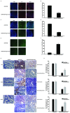
Figure 2. Wogonoside inhibited inflammatory cells infiltration and colon cancer progression in the tumor inflammatory microenvironment in vivo
A–B. The distribution of neutrophil (Gr-1+) and C–D. macrophage (F4/80+) infiltration were observed by confocal laser-scanning microscope. The results are representative of three independent experiments and expressed as mean ± SD, **P < 0.01 compared with AOM/DSS group. The results are representative of three independent experiments and expressed as mean ± SD, **P < 0.01 compared with AOM/DSS group. E–F. Immunofluorescent TUNEL staining was detected. G–H. Immunohistochemistry of Ki-67, BrdU and PCNA in surrounding and tumor tissues were shown with brown colored positive cells. The results are representative of three independent experiments and expressed as mean ± SD, *P < 0.05, **P < 0.01 compared with AOM/DSS group.
Figure 3
Figure 3. Wogonoside reduced production of pro-inflammatory cytokines and inhibited NF-κB activation via regulating PI3K/Akt pathway in AOM/DSS-induced adenocarcinoma
A–B. The expression of IL-1β, IL-6, TNF-α and NF-κB p65 in surrounding and tumor tissues of AOM/DSS-treated mice was performed by immunohistochemistry. The results are representative of three independent experiments and expressed as mean ± SD, *P < 0.05, **P < 0.01 compared with AOM/DSS group. C. The mRNA levels of IL-1β, IL-6 and TNF-α in tumor tissues. The results are representative of three independent experiments and expressed as mean ± SD, **P < 0.01 compared with AOM/DSS group. D. NF-kB p65 nuclear translocation in tumor tissues were determined by Western Blot. E. Densitometric analysis was performed to determine the relative ratios of each protein. Lamin A and β-actin were used as nuclear and cytoplasmic markers, respectively. The results are representative of three independent experiments and expressed as mean ± SD, ## P<0.01 compared with normal group; **P<0.01 compared with AOM/DSS group. F. The protein expression of p-p65 in tumor tissues. G. Densitometric analysis to determine the relative ratio normalized to β-actin. The results are representative of three independent experiments and expressed as mean ± SD, ## P<0.01 compared with normal group; **P<0.01compared with AOM/DSS group. H. The protein expression of PI3K, p-Akt, Akt, p-IKKα, IKKα, p-IκBα, IκBα, Cyclin D1 and survivin in tumor tissues was analyzed by Western Blot. I. Densitometric analysis to determine the relative ratio normalized to β-actin. The results are representative of three independent experiments and expressed as mean ± SD, ## P<0.01 compared with normal group; **P<0.01compared with AOM/DSS group.
Figure 4
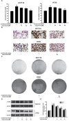
Figure 4. Wogonoside inhibited the growth of human colon cancer cells cultured with the conditioned media from LPS-activated THP-1 cells
A. MTT assays of HCT-116 and HT29 cells were performed to detect the inhibitory effect of wogonoside on the cell viability. The results are representative of three independent experiments and expressed as mean ± SD, ## P<0.01 compared with control group; *P<0.05, **P<0.01 compared with conditioned media group. B. Ki67 cell proliferation detection of HCT-116 and HT29 cells in conditional culture system treated with wogonoside. C. Soft-sugar-colony forming experiment was performed to ascertain the inhibitory effect of wogonoside on cell proliferation. D. The protein expression of PCNA was analyzed by Western Blot. E. Densitometric analysis to determine the relative ratio normalized to β-actin. The results are representative of three independent experiments and expressed as mean ± SD, ## P<0.01 compared with control group; **P<0.01 compared with conditioned media group.
Figure 5
Figure 5. Wogonoside inhibited the activation of NF-κB induced by the conditioned media from LPS-activated THP-1 cells in human colon cancer cells
A. The protein expression of p-IKKα, IKKα, p-IκBα, IκBα, Cyclin D1 and survivin in HCT116 and HT29 cells in all groups was analyzed by Western Blot. B. Densitometric analysis to determine the relative ratio normalized to β-actin. The results are representative of three independent experiments and expressed as mean ± SD. ##P < 0.01 compared with control group; *P < 0.05, **P < 0.01 compared with conditioned media group. C. NF-κB p65 nuclear translocation in HCT116 and HT29 cells in all group were determined by Western Blot. D. Densitometric analysis was performed to determine the relative ratios of each protein. Lamin A and β-actin were used as nuclear and cytoplasmic markers, respectively. The results are representative of three independent experiments and expressed as mean ± SD. ##P < 0.01 compared with control group; *P < 0.05, **P < 0.01 compared with conditioned media group. E–F. Immunofluorescence was performed to analyze NF-κB p65 nuclear translocation in HCT116 and HT29 cells. The results are representative of three independent experiments and expressed as mean ± SD. ##P < 0.01 compared with control group; **P < 0.01 compared with conditioned media group. G. The transcriptional activities of p-NF-κB p65 in HCT116 and HT29 cells were determined by Luciferase activity assay. The results are representative of three independent experiments and expressed as mean ± SD. ##P < 0.01 compared with control group; *P < 0.05, **P < 0.01 compared with conditioned media group.
Figure 6
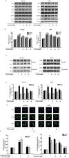
Figure 6. Wogonoside inhibited the proliferation of human colon cancer cells exposed to the conditioned media from LPS-activated THP-1 cells via inhibition of NF-κB activation through PI3K/Akt pathway
A. The protein expression of PI3K, p-Akt, Akt in HCT116 and HT29 cells in all groups was analyzed by Western Blot. B. Densitometric analysis to determine the relative ratio normalized to β-actin. The results are representative of three independent experiments and expressed as mean ± SD. ##P < 0.01 compared with control group; *P < 0.05, **P < 0.01 compared with conditioned media group. (C–H) HCT116 and HT29 cells were cultured in the conditional culture system with wogonoside (150 μM) or LY294002 (20 μM) for 24 h. C. Ki67 cell proliferation detection of HCT-116 and HT29 cells in the conditional culture system treated with wogonoside or LY294002. D. The protein expression of p-Akt, Akt, p-IKKα, IKKα, p-IκBα, IκBα and p-p65 was analyzed by Western Blot. E. Densitometric analysis to determine the relative ratio normalized to β-actin. The results are representative of three independent experiments and expressed as mean ± SD, ## P<0.01 compared with control group; **P<0.01 compared with conditioned media group. F. NF-κB p65 nuclear translocation in HCT116 and HT29 cells in all group were determined by Western Blot. G. Densitometric analysis was performed to determine the relative ratios of each protein. Lamin A and β-actin were used as nuclear and cytoplasmic markers, respectively. The results are representative of three independent experiments and expressed as mean ± SD. ##P < 0.01 compared with control group; **P < 0.01 compared with conditioned media group. H. The transcriptional activities of p-NF-κB p65 in HCT116 and HT29 cells in all groups were determined by Luciferase activity assay. The results are representative of three independent experiments and expressed as mean ± SD. ##P < 0.01 compared with control group; *P < 0.05, **P < 0.01 compared with conditioned media group. (I–M) HCT116 and HT29 cells were treated with IGF-1 (20 ng/ml) with or without wogonoside with for 24 h. I. Ki67 cell proliferation detection of HCT-116 and HT29 cells treated with IGF-1 with or without wogonoside. J. The protein expression of PI3K, p-Akt, Akt, p-IKKα, IKKα, p-IκBα and IκBα was analyzed by Western Blot. K. Densitometric analysis to determine the relative ratio normalized to β-actin. The results are representative of three independent experiments and expressed as mean ± SD, ## P<0.01 compared with control group; **P<0.01 compared with IGF-1 group. L. NF-κB p65 nuclear translocation in HCT116 and HT29 cells in all groups were determined by Western Blot. M. Densitometric analysis was performed to determine the relative ratios of each protein. Lamin A and β-actin were used as nuclear and cytoplasmic markers, respectively. The results are representative of three independent experiments and expressed as mean ± SD. ##P < 0.01 compared with control group; **P < 0.01 with IGF-1 group. N. The transcriptional activities of p-NF-κB p65 induced by IGF-1 in HCT116 and HT29 cells were determined by Luciferase activity assay. The results are representative of three independent experiments and expressed as mean ± SD. ##P < 0.01 compared with control group; **P < 0.01 with IGF-1 group.

Similar articles

Cited by

References

    1. Parkin DM, Bray F, Ferlay J, Pisani P. Global cancer statistics, 2002. CA Cancer J Clin. 2005;55:74–108. - PubMed
    1. Jemal A, Siegel R, Ward E, Hao Y, Xu J, Murray T, Thun MJ. Cancer statistics, 2008. CA Cancer J Clin. 2008;58:71–96. - PubMed
    1. Danese S, Mantovani A. Inflammatory bowel disease and intestinal cancer: a paradigm of the Yin-Yang interplay between inflammation and cancer. Oncogene. 2010;29:3313–3323. - PubMed
    1. Vakkila J, Lotze MT. Inflammation and necrosis promote tumour growth. Nat Rev Immunol. 2004;4:641–648. - PubMed
    1. Balkwill F, Mantovani A. Inflammation and cancer: back to Virchow? Lancet. 2001;357:539–545. - PubMed

MeSH terms