. 2016 Jun;5(6):836-44.
doi: 10.5966/sctm.2015-0158. Epub 2016 Apr 21.

Local Application of Isogenic Adipose-Derived Stem Cells Restores Bone Healing Capacity in a Type 2 Diabetes Model

Affiliations

Local Application of Isogenic Adipose-Derived Stem Cells Restores Bone Healing Capacity in a Type 2 Diabetes Model

Christoph Wallner et al. Stem Cells Transl Med. 2016 Jun.

Abstract

Bone regeneration is typically a reliable process without scar formation. The endocrine disease type 2 diabetes prolongs and impairs this healing process. In a previous work, we showed that angiogenesis and osteogenesis-essential steps of bone regeneration-are deteriorated, accompanied by reduced proliferation in type 2 diabetic bone regeneration. The aim of the study was to improve these mechanisms by local application of adipose-derived stem cells (ASCs) and facilitate bone regeneration in impaired diabetic bone regeneration. The availability of ASCs in great numbers and the relative ease of harvest offers unique advantages over other mesenchymal stem cell entities. A previously described unicortical tibial defect model was utilized in diabetic mice (Lepr(db-/-)). Isogenic mouse adipose-derived stem cells (mASCs)(db-/db-) were harvested, transfected with a green fluorescent protein vector, and isografted into tibial defects (150,000 living cells per defect). Alternatively, control groups were treated with Dulbecco's modified Eagle's medium or mASCs(WT). In addition, wild-type mice were identically treated. By means of immunohistochemistry, proteins specific for angiogenesis, cell proliferation, cell differentiation, and bone formation were analyzed at early (3 days) and late (7 days) stages of bone regeneration. Additionally, histomorphometry was performed to examine bone formation rate and remodeling. Histomorphometry revealed significantly increased bone formation in mASC(db-/db-)-treated diabetic mice as compared with the respective control groups. Furthermore, locally applied mASCs(db-/db-) significantly enhanced neovascularization and osteogenic differentiation. Moreover, bone remodeling was upregulated in stem cell treatment groups. Local application of mACSs can restore impaired diabetic bone regeneration and may represent a therapeutic option for the future.

Significance: This study showed that stem cells obtained from fat pads of type 2 diabetic mice are capable of reconstituting impaired bone regeneration in type 2 diabetes. These multipotent stem cells promote both angiogenesis and osteogenesis in type 2 diabetic bony defects. These data might prove to have great clinical implications for bony defects in the ever-increasing type 2 diabetic patient population.

Keywords: Bone; Diabetes mellitus; Fractures; Osteogenesis; Stem cells.


Figures

Figure 1.
Figure 1.
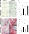
mASCsdb−/db− showed increased osteogenic differentiation as compared with mASCsWT. (A): Alkaline phosphatase (ALP) activity on 7 days differentiated mASCs from diabetic and WT animals in both media (5.5 and 22 mM glucose). mASCs from diabetic origin showed increased activity. (B): Photometric measurement of ALP reaction. (C): Alizarin red staining of osteogenically differentiated mASCs in normoglycemic and hyperglycemic medium (5.5 and 22 mM glucose) showed increased mineralization as compared with mASCsWT in both media. (D): Quantification of Alizarin red-positive pixels revealed highly significant increased mineralization rates of mASCsdb−/db− in both media compared with mASCsWT . Results are shown as means ± SEM. ∗, p < .05; ∗∗, p < .01; ∗∗∗, p < .001 (two-sample t test). Scale bars = 10 μm (A), 15 μm (C). Abbreviations: a.u., arbitrary units; mASC, mouse adipose-derived stem cell; WT, wild type.
Figure 2.
Figure 2.
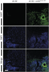
Successful colonization of mASCsdb−/db−GFP+ in diabetic defects. Defects were colonized with isogenic GFP-positive (green) mASCsdb−/db− (1.5 × 10⁵ mASCsdb−/db−; right column). GFP-positive cells indicate successful colonization (left column; comparison with diabetic defect without treatment). Scale bar = 20 μm. Abbreviations: DAPI, 4′,6-diamidino-2-phenylindole; EGFP, enhanced green fluorescent protein; GFP, green fluorescent protein; mASC, mouse adipose-derived stem cell.
Figure 3.
Figure 3.
Isogenic mASCsdb−/db− and mASCWT increase angiogenesis in diabetic defects. (A): Blood vessels and endothelial cells were detected by immunofluorescence for PECAM-1 (3 dpo). db/db control mice exhibited decreased angiogenesis compared with control WT mice (left column), whereas defects treated with mASCsdb−/db− (right column) showed a significant improvement in angiogenesis compared with control animals without treatment (db-Ctrl). Although all mASC-transplanted mice exhibited improved angiogenesis, mASCsdb−/db− did show the highest increase in angiogenesis. (B): Higher magnifications from A (white box). (C): Quantification of PECAM-1-positive pixels (arbitrary units) revealed that angiogenesis is significantly increased in mASCs-treated defects compared with diabetic defects without treatment and rescued to levels of WT control mice. mASCs transplanted into WT mice have shown a further improvement to WT-control animals. (D): Blood vessels and endothelial cells were detected by immunofluorescence for VCAM-1 (3 dpo). Results largely paralleled PECAM-1 staining. (E): Higher magnifications from (D) (white box). (F): Quantification of VCAM-1-positive pixels (arbitrary units) revealed that angiogenesis is significantly increased in mASC-treated defects compared with diabetic defects without treatment and rescued to levels of WT mice. Results are shown as means ± SEM. ∗∗, p < .01; ∗∗∗, p < .001 (two-sample t test). Scale bars = 200 μm (A, D), 20 μm (B, E). Abbreviations: a.u., arbitrary units; Ctrl, control; dpo, days postoperation; GFP, green fluorescent protein; mASC, mouse adipose-derived stem cell; PECAM-1, platelet endothelial cell adhesion molecule 1; VCAM-1, vascular cell adhesion molecule 1; WT, wild type.
Figure 4.
Figure 4.
mASCsdb−/db− application enhances proliferation and differentiation in diabetic bony defects. (A): Immunohistochemistry for PCNA, RUNX-2, and osteocalcin revealed enhancement of osteogenic and proliferative capacity in mASCsdb−/db−-treated diabetic animals compared with control group (db/db). (B): Quantification of Nova Red-positive pixels showed that cell proliferation and differentiation are significantly increased in diabetic animals treated with mASCsdb−/db− compared with untreated animals. Application of mASCsWT into diabetic defects showed no significant improvement in PCNA, RUNX-2, or osteocalcin expression. WT defects treated with mASCs showed no significant alteration in PCNA, RUNX-2, or osteocalcin expression. Results are shown as means ± SEM. ∗, p < .05; ∗∗, p < .01 (two-sample t test). Scale bar = 200 μm. Abbreviations: Ctrl, control; dpo, days postoperation; mASC, mouse adipose-derived stem cell; PCNA, proliferating cell nuclear antigen; RUNX-2, Runt-related transcription factor 2; WT, wild type.
Figure 5.
Figure 5.
mASCsdb−/db− increase bone regeneration in diabetic bony defects. (A): Aniline blue staining of db/db control mice showed impaired new bone formation compared with WT control mice (left column), whereas diabetic defects treated with mASCsdb−/db− showed improved bone formation (right column) (7 dpo). Transplantation of mASCsdb−/db− and mASCsWT into WT mice did not improve bone formation. (B): Extracted osteoid formation area. (C): Quantification of aniline blue-positive pixels revealed that bone formation is significantly increased in mASC-treated diabetic defects compared with the control group db/db and similar to bone formation of WT mice. mASC implantation into WT mice did not show improved bone regeneration. Results are shown as means ± SEM. Scale bar = 200 μm. ∗, p < .05; ∗∗, p < .01; ∗∗∗, p < .001 (two-sample t test). Abbreviations: Cb, cortical bone; Ctrl, control; d, days postoperation; mASC, mouse adipose-derived stem cell; n.s., not significant; WT, wild type.
Figure 6.
Figure 6.
Paracrine effects dominate increases in angiogenic and osteogenic differentiation mediated by mASCsdb−/db−GFP+ Immunofluorescence stainings were performed for PECAM, RUNX-2, and PCNA in diabetic mice transplanted with mASCdb−/db−GFP+ and controls. EGFP-positive areas indicate transplanted stem cells. Scale bar = 40 μm. Abbreviations: DAPI, 4′,6-diamidino-2-phenylindole; EGFP, enhanced green fluorescent protein; GFP, green fluorescent protein; mASC, mouse adipose-derived stem cell; PCNA, proliferating cell nuclear antigen; PECAM, platelet endothelial cell adhesion molecule; RUNX-2, Runt-related transcription factor 2.

References

    1. Gaston MS, Simpson AHRW. Inhibition of fracture healing. J Bone Joint Surg Br. 2007;89-B:1553–1560. - PubMed
    1. Retzepi M, Donos N. The effect of diabetes mellitus on osseous healing. Clin Oral Implants Res. 2010;21:673–681. - PubMed
    1. Daneman D. Type 1 diabetes. Lancet. 2006;367:847–858. - PubMed
    1. Wild S, Roglic G, Green A, et al. Global prevalence of diabetes: eEstimates for the year 2000 and projections for 2030. Diabetes Care. 2004;27:1047–1053. - PubMed
    1. Isidro ML, Ruano B. Bone disease in diabetes. Curr Diabetes Rev. 2010;6:144–155. - PubMed

Publication types