

Isolation of Pancreatic Cancer Cells from a Patient-Derived Xenograft Model Allows for Practical Expansion and Preserved Heterogeneity in Culture
- PMID: 27102771
- PMCID: PMC4901138
- DOI: 10.1016/j.ajpath.2016.02.009
Isolation of Pancreatic Cancer Cells from a Patient-Derived Xenograft Model Allows for Practical Expansion and Preserved Heterogeneity in Culture
Abstract
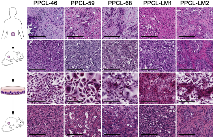
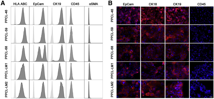
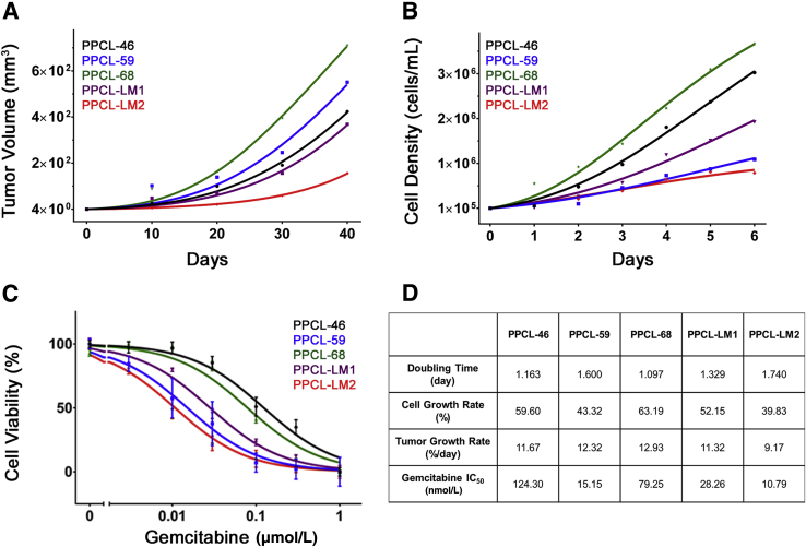
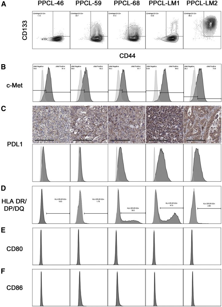
Commercially available, highly passaged pancreatic cancer (PC) cell lines are of limited translational value. Attempts to overcome this limitation have primarily consisted of cancer cell isolation and culture directly from human PC specimens. However, these techniques are associated with exceedingly low success rates. Here, we demonstrate a highly reproducible culture of primary PC cell lines (PPCLs) from patient-derived xenografts, which preserve, in part, the intratumoral heterogeneity known to exist in PC. PPCL expansion from patient-derived xenografts was successful in 100% of attempts (5 of 5). Phenotypic analysis was evaluated with flow cytometry, immunofluorescence microscopy, and short tandem repeat profiling. Importantly, tumorigenicity of PPCLs expanded from patient-derived xenografts was assessed by subcutaneous injection into nonobese diabeteic.Cg-Prkdc(scid)Il2rg(tm1Wjl)/SzJ mice. Morphologically, subcutaneous injection of all PPCLs into mice yielded tumors with similar characteristics to the parent xenograft. PPCLs uniformly expressed class I human leukocyte antigen, epithelial cell adhesion molecule, and cytokeratin-19. Heterogeneity within each PPCL persisted in culture for the frequency of cells expressing the cancer stem cell markers CD44, CD133, and c-Met and the immunologic markers human leukocyte antigen class II and programmed death ligand 1. This work therefore presents a reliable method for the rapid expansion of primary human PC cells and, thereby, provides a platform for translational investigation and, importantly, potential personalized therapeutic approaches.
Copyright © 2016 American Society for Investigative Pathology. Published by Elsevier Inc. All rights reserved.
Figures
Similar articles
-
Patient-derived xenografts of gastrointestinal cancers are susceptible to rapid and delayed B-lymphoproliferation.Int J Cancer. 2017 Mar 15;140(6):1356-1363. doi: 10.1002/ijc.30561. Int J Cancer. 2017. PMID: 27935045
-
Orthotopic Patient-Derived Pancreatic Cancer Xenografts Engraft Into the Pancreatic Parenchyma, Metastasize, and Induce Muscle Wasting to Recapitulate the Human Disease.Pancreas. 2017 Jul;46(6):813-819. doi: 10.1097/MPA.0000000000000843. Pancreas. 2017. PMID: 28609371 Free PMC article.
-
Esophageal Adenocarcinoma Cells and Xenograft Tumors Exposed to Erb-b2 Receptor Tyrosine Kinase 2 and 3 Inhibitors Activate Transforming Growth Factor Beta Signaling, Which Induces Epithelial to Mesenchymal Transition.Gastroenterology. 2017 Jul;153(1):63-76.e14. doi: 10.1053/j.gastro.2017.03.004. Epub 2017 Mar 9. Gastroenterology. 2017. PMID: 28286209
-
Patient-derived xenograft models for pancreatic adenocarcinoma demonstrate retention of tumor morphology through incorporation of murine stromal elements.Am J Pathol. 2015 May;185(5):1297-303. doi: 10.1016/j.ajpath.2015.01.016. Epub 2015 Mar 12. Am J Pathol. 2015. PMID: 25770474 Free PMC article.
-
Phenotypic characterization of human colorectal cancer stem cells.Proc Natl Acad Sci U S A. 2007 Jun 12;104(24):10158-63. doi: 10.1073/pnas.0703478104. Epub 2007 Jun 4. Proc Natl Acad Sci U S A. 2007. PMID: 17548814 Free PMC article.
Cited by
-
The crosstalk between macrophages and cancer cells potentiates pancreatic cancer cachexia.Cancer Cell. 2024 May 13;42(5):885-903.e4. doi: 10.1016/j.ccell.2024.03.009. Epub 2024 Apr 11. Cancer Cell. 2024. PMID: 38608702 Free PMC article.
-
Tumor-intrinsic PIK3CA represses tumor immunogenecity in a model of pancreatic cancer.J Clin Invest. 2019 May 21;129(8):3264-3276. doi: 10.1172/JCI123540. J Clin Invest. 2019. PMID: 31112530 Free PMC article.
-
The role of survivin in the progression of pancreatic ductal adenocarcinoma (PDAC) and a novel survivin-targeted therapeutic for PDAC.PLoS One. 2020 Jan 13;15(1):e0226917. doi: 10.1371/journal.pone.0226917. eCollection 2020. PLoS One. 2020. PMID: 31929540 Free PMC article.
-
IL-8 Released from Human Pancreatic Cancer and Tumor-Associated Stromal Cells Signals through a CXCR2-ERK1/2 Axis to Induce Muscle Atrophy.Cancers (Basel). 2019 Nov 25;11(12):1863. doi: 10.3390/cancers11121863. Cancers (Basel). 2019. PMID: 31769424 Free PMC article.
-
Comparative study on contrast enhancement of Magnevist and Magnevist-loaded nanoparticles in pancreatic cancer PDX model monitored by MRI.Cancer Nanotechnol. 2020;11:5. doi: 10.1186/s12645-020-00061-9. Epub 2020 May 14. Cancer Nanotechnol. 2020. PMID: 32714466 Free PMC article.
References
-
- Rahib L., Smith B.D., Aizenberg R., Rosenzweig A.B., Fleshman J.M., Matrisian L.M. Projecting cancer incidence and deaths to 2030: the unexpected burden of thyroid, liver, and pancreas cancers in the United States. Cancer Res. 2014;74:2913–2921. - PubMed
-
- Koay E.J., Truty M.J., Cristini V., Thomas R.M., Chen R., Chatterjee D., Kang Y., Bhosale P.R., Tamm E.P., Crane C.H., Javle M., Katz M.H., Gottumukkala V.N., Rozner M.A., Shen H., Lee J.E., Wang H., Chen Y., Plunkett W., Abbruzzese J.L., Wolff R.A., Varadhachary G.R., Ferrari M., Fleming J.B. Transport properties of pancreatic cancer describe gemcitabine delivery and response. J Clin Invest. 2014;124:1525–1536. - PMC - PubMed
-
- Moore M.J., Goldstein D., Hamm J., Figer A., Hecht J.R., Gallinger S., Au H.J., Murawa P., Walde D., Wolff R.A., Campos D., Lim R., Ding K., Clark G., Voskoglou-Nomikos T., Ptasynski M., Parulekar W. Erlotinib plus gemcitabine compared with gemcitabine alone in patients with advanced pancreatic cancer: a phase III trial of the National Cancer Institute of Canada Clinical Trials Group. J Clin Oncol. 2007;25:1960–1966. - PubMed
-
- Baxter N.N., Whitson B.A., Tuttle T.M. Trends in the treatment and outcome of pancreatic cancer in the United States. Ann Surg Oncol. 2007;14:1320–1326. - PubMed
-
- Yunis A.A., Arimura G.K., Russin D.J. Human pancreatic carcinoma (MIA PaCa-2) in continuous culture: sensitivity to asparaginase. Int J Cancer. 1977;19:128–135. - PubMed
Publication types
MeSH terms
Grants and funding
LinkOut - more resources
Full Text Sources
Other Literature Sources
Medical
Research Materials
Miscellaneous
