. 2016 Apr 22:6:24922.
doi: 10.1038/srep24922.

Long non-coding RNAs harboring miRNA seed regions are enriched in prostate cancer exosomes

Affiliations

Long non-coding RNAs harboring miRNA seed regions are enriched in prostate cancer exosomes

Alireza Ahadi et al. Sci Rep. .

Abstract

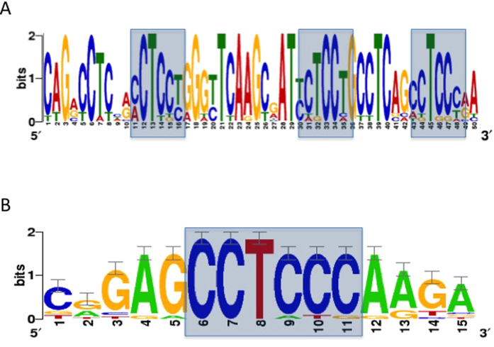
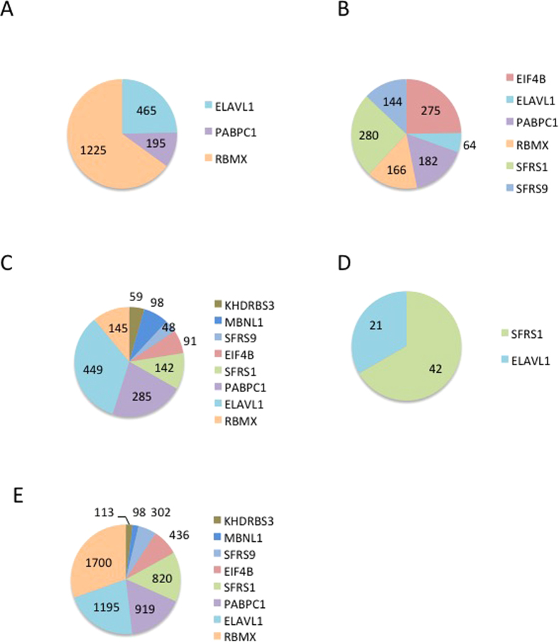
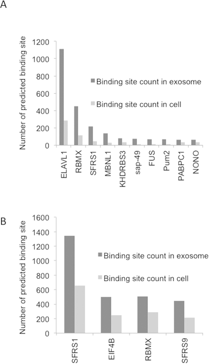
Long non-coding RNAs (lncRNAs) form the largest transcript class in the human transcriptome. These lncRNA are expressed not only in the cells, but they are also present in the cell-derived extracellular vesicles such as exosomes. The function of these lncRNAs in cancer biology is not entirely clear, but they appear to be modulators of gene expression. In this study, we characterize the expression of lncRNAs in several prostate cancer exosomes and their parental cell lines. We show that certain lncRNAs are enriched in cancer exosomes with the overall expression signatures varying across cell lines. These exosomal lncRNAs are themselves enriched for miRNA seeds with a preference for let-7 family members as well as miR-17, miR-18a, miR-20a, miR-93 and miR-106b. The enrichment of miRNA seed regions in exosomal lncRNAs is matched with a concomitant high expression of the same miRNA. In addition, the exosomal lncRNAs also showed an over representation of RNA binding protein binding motifs. The two most common motifs belonged to ELAVL1 and RBMX. Given the enrichment of miRNA and RBP sites on exosomal lncRNAs, their interplay may suggest a possible function in prostate cancer carcinogenesis.


Figures

Figure 1
Figure 1. Characterisation of exosomes from in vitro cell culture.
(A) Representative TEM image of DU145 exosomes at 7900 magnifications. White arrows depict exosomes of ~100 μm with kidney bean like appearances. (B) Representative particle tracking of a DU145 exosomes using the NanoSight Instrumentation. (C) Detection of exosome surface markers, CD9, CD63, TSG101 and AGO2 in DU145 and PNT2 cells. Absorbance was measured by the release of europium using a modified ELISA. (D) Small RNA bioanalyser chip image showing the presences of small ncRNAs between 21 and 27 nts (black arrows).
Figure 2
Figure 2
(A) Venn diagram showing the common exosomal lncRNAs between the PC3, DU145, LNCaP and VCaP prostate cancer cell lines. (B) Heat map showing the expression of exosomal lncRNAs in prostate cancer cells and a normal cell line (PNT2). Red denotes high expression and aqua denotes reduced expression of the lncRNAs.
Figure 3
Figure 3
(A) miRNA heatmap in exosomes representing prostate cancer and PNT2 normal cell lines. Aqua colour represents reduced levels and Red is indicative of high expression. (B) Differential expression of miRNAs in the parental five cell lines. (C) Heatmap showing the expression of miRNA in both exosomes and their parental cell line.
Figure 4
Figure 4
Gapped local alignments of motifs in (A) exosomal lncRNAs. (B) exosomal lncRNAs vs cellular lncRNAs (control).
Figure 5
Figure 5
The number of positive matching RBP binding sites found in the exosomal lncRNAs for each cell line (A) VCaP, (B) PC3, (C) LNCaP, (D) DU145, (E) Distribution of RNA binding sites in exosomal lncRNAs of at least one cancer cell line.
Figure 6
Figure 6. Comparison of RNA binding motifs on exosomal lncRNAs versus cellular transcripts.
(A) Reduction of RBP motifs in cellular lncRNAs which are common to all four cell lines. In contrast there is a increase of these motifs in the exosomal lncRNAs. (B) Increase in RBP motifs of the enriched exosomal lncRNAs (141 transcripts) to those lncRNAs, which were highly expressed in the cells (151 transcripts) of at least one cell line. This analysis yielded four major binding sites for, RBMX, SFRS1, SFRS9 and EIF4B.

References

    1. Mattick J. S. & Makunin I. V. Non-coding RNA. Hum Mol Genet 15 Spec No 1, R17–29, doi: 10.1093/hmg/ddl046 (2006). - DOI - PubMed
    1. Jacquier A. The complex eukaryotic transcriptome: unexpected pervasive transcription and novel small RNAs. Nat Rev Genet 10, 833–844, doi: 10.1038/nrg2683 (2009). - DOI - PubMed
    1. Carninci P. et al. The transcriptional landscape of the mammalian genome. Science 309, 1559–1563, doi: 10.1126/science.1112014 (2005). - DOI - PubMed
    1. Johnson J. M., Edwards S., Shoemaker D. & Schadt E. E. Dark matter in the genome: evidence of widespread transcription detected by microarray tiling experiments. Trends Genet 21, 93–102, doi: 10.1016/j.tig.2004.12.009 (2005). - DOI - PubMed
    1. Guffanti A. et al. A transcriptional sketch of a primary human breast cancer by 454 deep sequencing. BMC Genomics 10, 163, doi: 10.1186/1471-2164-10-163 (2009). - DOI - PMC - PubMed