. 2016 Apr 22:6:24629.
doi: 10.1038/srep24629.

Simultaneous hyperthermia-chemotherapy with controlled drug delivery using single-drug nanoparticles

Affiliations

Simultaneous hyperthermia-chemotherapy with controlled drug delivery using single-drug nanoparticles

Itaru Sato et al. Sci Rep. .

Abstract

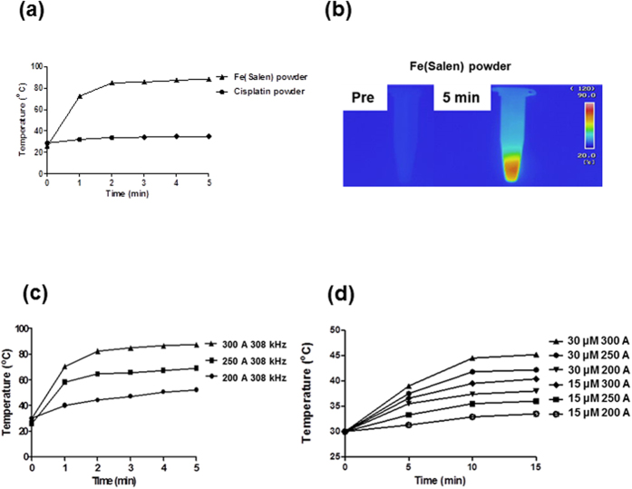
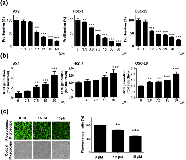
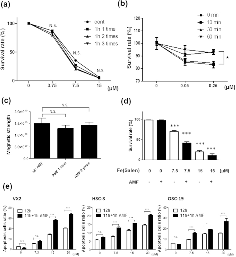
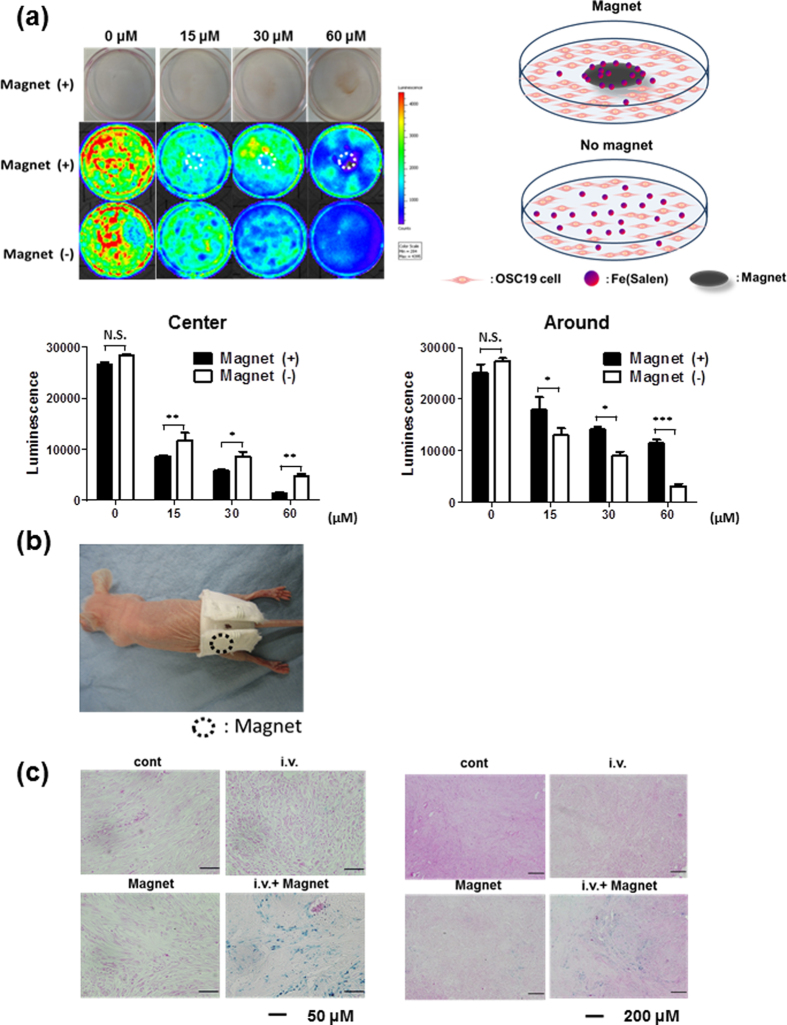
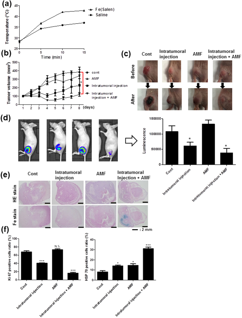
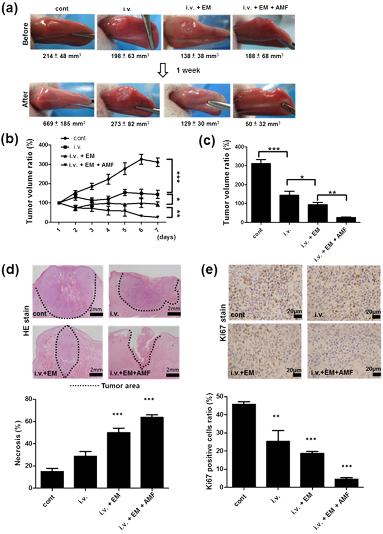
We previously investigated the utility of μ-oxo N,N'- bis(salicylidene)ethylenediamine iron (Fe(Salen)) nanoparticles as a new anti-cancer agent for magnet-guided delivery with anti-cancer activity. Fe(Salen) nanoparticles should rapidly heat up in an alternating magnetic field (AMF), and we hypothesized that these single-drug nanoparticles would be effective for combined hyperthermia-chemotherapy. Conventional hyperthermic particles are usually made of iron oxide, and thus cannot exhibit anti-cancer activity in the absence of an AMF. We found that Fe(Salen) nanoparticles induced apoptosis in cultured cancer cells, and that AMF exposure enhanced the apoptotic effect. Therefore, we evaluated the combined three-fold strategy, i.e., chemotherapy with Fe(Salen) nanoparticles, magnetically guided delivery of the nanoparticles to the tumor, and AMF-induced heating of the nanoparticles to induce local hyperthermia, in a rabbit model of tongue cancer. Intravenous administration of Fe(Salen) nanoparticles per se inhibited tumor growth before the other two modalities were applied. This inhibition was enhanced when a magnet was used to accumulate Fe(Salen) nanoparticles at the tongue. When an AMF was further applied (magnet-guided chemotherapy plus hyperthermia), the tumor masses were dramatically reduced. These results indicate that our strategy of combined hyperthermia-chemotherapy using Fe(Salen) nanoparticles specifically delivered with magnetic guidance represents a powerful new approach for cancer treatment.


Figures

Figure 1
Figure 1. Heat is generated by Fe(Salen) nanoparticles upon exposure to an AMF.
(a) Heat generation by cisplatin or Fe(Salen) upon AMF exposure. The AMF was applied at a frequency of 308 kHz and EC 250 A. (b) Representative thermography of Fe(Salen) nanoparticle dry powder in a tube before (Pre) and 5 min after AMF exposure (5 min). (c) Effect of an AMF (200, 250 and 300 A) on the temperature of Fe(Salen) nanoparticles in culture medium. (d) Effect of Fe(Salen) concentration (15 or 30 μM) and electrical current (200, 250 or 300 A) on the temperature in the culture medium.
Figure 2
Figure 2. Fe(Salen) nanoparticles inhibit cell proliferation, promote ROS generation, and are taken up by cells.
(a) Effect of Fe(Salen) on proliferation of various cancer cells. XTT cell proliferation assays were performed with human and rabbit tongue cancer cells: VX2 rabbit squamous cell carcinoma, HSC-3 human squamous cell carcinoma, and OSC-19 human squamous cell carcinoma (n = 4). The IC50 values were similar among cell types and were approximately 7.5 μM. (b) Effect of Fe(Salen) on ROS production in various cells. Fe(Salen) nanoparticles generated ROS in a concentration-dependent manner (n = 4, **p < 0.01, ***p < 0.001). (c) Representative fluorescence pictures of calcein using a fluorescence microscope and optical microscope. Ratios of calcein fluorescence are shown below (n = 4, **p < 0.01, ***p < 0.001 vs. control). Note that cellular fluorescence was decreased in the presence of Fe(Salen) nanoparticles.
Figure 3
Figure 3. Fe(Salen) nanoparticle-induced apoptosis is increased by AMF exposure.
(a) Effect of an AMF on Fe(Salen) nanoparticles. Fe(Salen) nanoparticles were heated to 80 °C by exposure to an AMF for 60 minutes once, twice, or three times. Changes in cytotoxic potency were examined in the presence of various concentrations of Fe(Salen) nanoparticles in VX2 cells. Note that there were no changes in cytotoxicity (n = 4, N.S., not significant). (b) Effect of high temperature (50 °C) on cytotoxic potency of Cetuximab (Erbitax®). Centuximab, a drug targeting epidermal growth factor receptor (EGFR), was heated to 50 °C for 30 or 60 minutes, followed by cytotoxicity assay in OSC-19 cells. Note that AMF exposure did not change the cytotoxicity of Fe(Salen) nanoparticles, but did change that of Cetuximab (Erbitax®) (n = 4, *p < 0.05). (c) ESR analysis of magnetism after exposure to an AMF. No AMF; Fe(Salen) without AMF exposure, AMF 1 time; Fe(Salen) with AMF exposure for an hour once, AMF 2 times; Fe(Salen) with AMF exposure for one hour twice. (d) Increased anti-cancer effect of Fe(Salen) with AMF exposure. An AMF promoted cellular death of VX2 cells in the presence of Fe(Salen) nanoparticles, as determined by trypan blue staining (n = 4, **p < 0.01, ***p < 0.001 vs. control). (e) Effect of Fe(Salen) with AMF exposure on various cancer cells. Apoptotic cell ratio is shown after incubation in the presence of various concentrations (0, 7.5, 15, 30 μM) of Fe(Salen) nanoparticles for 12 hours (white bars) or for 11 hours followed by 1 hour AMF exposure (black bars). Apoptosis was determined by Annexin-V/PI staining with FACS scan dot plot analysis at 12 hours after treatment with Fe(Salen) nanoparticles. Note that AMF exposure increased the cytotoxicity in the presence of Fe(Salen) nanoparticles (n = 4, *p < 0.05).
Figure 4
Figure 4. Fe(Salen) nanoparticles are attracted by a permanent magnet in vitro and in the mouse model in vivo.
(a) Distribution of Fe(Salen) and cellular death in the presence of a magnet. Distributions of Fe(Salen) and cytotoxic effect were compared between the center (center), where the magnet was positioned, and the edge (edge) of each culture dish. Upper photo: distribution of various concentrations of Fe(Salen) in a dish with a magnet. Middle photo: Cell viability in the presence of a magnet. Cell viability was determined in terms of luciferase activity by intensity measurement with an IVIS imaging system. Lower photo: Cell viability in the absence of a magnet. Bar graphs show the determination of cell viability with IVIS (center and edge). (n = 4, ***p < 0.001) Note that the cytotoxicity of Fe(Salen) nanoparticles was enhanced in the center. In contrast, the cell viability at the edge of culture dishes is maintained in the presence of a magnet compared to that of in the absence of a magnet. (b) A Jacket used for drug delivery in mice. Circle shows the site where the permanent magnet is installed. (c) Representative photo of skin tissues at the site beneath the magnet in mice which had been injected with Fe(Salen) via the tail vein. Fe(Salen) was accumulated beneath the magnet at the tumor location. Mice were intravenously injected with Fe(Salen) (5 mg/kg) and wore a jacket for three days. Accumulation of Fe(Salen) nanoparticles was examined by Berlin blue staining. Cont; control, iv; Fe(Salen) was injected, Magnet; jacket was worn, iv Magnet; Fe(Salen) was injected and the jacket was worn. High (left, calibration bar 50 μm) and low magnification (right, 200 μm) images are shown.
Figure 5
Figure 5. Local injection of Fe(Salen) nanoparticles generates heat upon exposure to an AMF; induced anti-cancer effect in a mouse tongue cancer model.
(a) Changes in temperature with AMF exposure. Either Fe(Salen) nanoparticles or saline were injected into the tumor in mice, followed by exposure to an AMF. Note that the local temperature was increased to a greater degree with Fe(Salen) in a time-dependent manner. (b) Effect of Fe(Salen) nanoparticle injection and AMF exposure on tumor size. The graph showed the time course of tumor volume changes. Fe(Salen) inhibited tumor growth, and AMF exposure of Fe(Salen)-injected animals further inhibited the tumor growth. Mean tumor volume (mm3) of each group is also shown. Control (cont), intratumoral injection of Fe(Salen) nanoparticles (injection), AMF exposure alone (AMF), and intratumor injection of Fe(Salen) nanoparticles and AMF exposure (injection and AMF). (n = 6, *p < 0.05, **p < 0.01, ***p < 0.001). (c) Representative photo of tumors in each group. (d) Effect of Fe(Salen) nanoparticle injection and AMP exposure on tumor size, determined from IVIS images. Representative IVIS images of mouse tumors (left) and luminescence intensity in each group (right) are shown. (e) Representative photos of tumor tissues from each group. HE staining (upper) and Fe staining (lower) are shown. Calibration bar: 2 mm. (f) Expression of Ki67-positive cells (left) and HSP70-positive cells (right).
Figure 6
Figure 6. Anti-cancer effect of Fe(Salen) nanoparticles with magnet application and AMF exposure in rabbits.
(a) Representative photos of rabbit tongue tumors in each group before (upper) and after (lower) treatment. Mean tumor volume value (mm3) of each group is also shown. Control (cont), intravenous injection of Fe(Salen) nanoparticles (i.v.), Fe(Salen) nanoparticle injection and electromagnet application (i.v. + DDS), and Fe(Salen) nanoparticle injection, electromagnet application, and AMF exposure (i.v. + DDS + AMF). (b) Changes in tumor volume ratio for 7 days. (n = 6, *p < 0.05, **p < 0.01, ***p < 0.001). (c) Comparison of tumor volume ratios at day 7 (n = 6, *p < 0.05, **p < 0.01,***p < 0.001). (d) Representative histological photo by HE staining at day 7 (upper). Broken lines indicate tumor areas. Calibration bar: 2 mm. Quantification of necrotic area by HE staining (lower) (n = 4, ***p < 0.001). (e) Representative pictures of tongue tumors after Ki67 staining (upper). Calibration bar: 20 μm. Quantification of necrosis by Ki67 staining in rabbit tongue tumors (lower) (n = 4, **p < 0.01, ***p < 0.001).

References

    1. Aguzzi A. et al. MAP kinase modulation in squamous cell carcinoma of the oral cavity. Anticancer Res 29, 303–308 (2009). - PubMed
    1. Tanaka T., Tanaka M. & Tanaka T. Oral carcinogenesis and oral cancer chemoprevention: A Review. Patholog Res Int 2011, 10 (2011). - PMC - PubMed
    1. Radiofrequency Hyperthermia for Cancer. Lancet 323, 885–886 (1984). - PubMed
    1. Suit H. D. & Shwayder M. Hyperthermia: Potential as an anti-tumor agent. Cancer 34, 122–129 (1974). - PubMed
    1. Abe M. H. et al. Multi-institutional studies on hyperthermia using an 8-MHz radiofrequency capacitive heating device (Thermotron RF-8) in combination with radiation for cancer therapy. Cancer 58, 1589–1595 (1986). - PubMed

Publication types

Substances