. 2016 May:41:25-38.
doi: 10.1016/j.neurobiolaging.2016.02.004. Epub 2016 Feb 12.

Aging is associated with dimerization and inactivation of the brain-enriched tyrosine phosphatase STEP

Affiliations

Aging is associated with dimerization and inactivation of the brain-enriched tyrosine phosphatase STEP

Sathyanarayanan Rajagopal et al. Neurobiol Aging. 2016 May.

Abstract

The STriatal-Enriched tyrosine Phosphatase (STEP) is involved in the etiology of several age-associated neurologic disorders linked to oxidative stress and is also known to play a role in neuroprotection by modulating glutamatergic transmission. However, the possible effect of aging on STEP level and activity in the brain is still unclear. In this study, using young (1 month), adult (4 months), and aged (18 months) rats, we show that aging is associated with increase in dimerization and loss of activity of STEP. Increased dimerization of STEP is primarily observed in the cortex and hippocampus and is associated with depletion of both reduced and total glutathione levels, suggesting an increase in oxidative stress. Consistent with this interpretation, studies in cell culture models of glutathione depletion and oxidative stress also demonstrate formation of dimers and higher order oligomers of STEP that involve intermolecular disulfide bond formation between multiple cysteine residues. Conversely, administration of N-acetyl cysteine, a major antioxidant that enhances glutathione biosynthesis, attenuates STEP dimerization both in the cortex and hippocampus. The findings indicate that loss of this intrinsic protective response pathway with age-dependent increase in oxidative stress may be a contributing factor for the susceptibility of the brain to age-associated neurologic disorders.

Keywords: Aging; Dimerization; Glutathione; N-acetyl cysteine; STEP; Tyrosine phosphatase.


Conflict of interest statement

Disclosure statement for authors

The authors declare that they have no conflicts of interest.

Figures

Figure 1
Figure 1
Aging leads to increased dimerization of STEP in cortex and hippocampus. Equal amounts of protein from (A) cortical, (B) hippocampal and (C) striatal lysates obtained from 1, 4 and 18 month old male SD rats were processed for gel electrophoresis under non-reducing conditions (without β-mercaptoethanol) followed by immunoblotting with anti-STEP antibody to evaluate STEP dimer formation (upper panel). Equal amounts of protein from the same samples were processed under reducing conditions (with β-mercaptoethanol) to assess STEP expression level (middle panel) and the blots were re-probed with anti-tubulin antibody to indicate equal protein loading (lower panel). (A–C) Quantification of dimeric STEP61 in non-reducing gels (band at 100–150 kDa) was performed by computer-assisted densitometry and image J analysis (right column). Values are mean ± SEM (n = 5). *p < 0.05, **p < 0.01.
Figure 2
Figure 2
Aging is associated with depletion of both reduced and total GSH levels. (A, D) Cortical, (B, E) hippocampal and (C, F) striatal lysates obtained from 1, 4 and 18 month old SD rats were processed for quantification of both reduced (A–C) and total (D–F) GSH levels. The fluorescent signal generated from the Thiostar-GSH adduct was measured fluorometrically (Ex/Em=390/510 nm). Bar diagrams represent mean ± SEM of reduced and total GSH levels (n = 5). *p < 0.05; **p < 0.005; ***p < 0.0001.
Figure 3
Figure 3
Aging is associated with decrease in STEP phosphatase activity. (A) Cortical, (B) hippocampal and (C) striatal lysates obtained from 1 and 18 month old SD rats were processed for immunoprecipitation of STEP using anti-STEP antibody. PTP activity was assayed using pNPP as a substrate. Quantitative measurement of the formation of para-nitrophenolate is represented as mean ± SEM (n = 4). *p < 0.01. In a parallel series of experiments, immunoprecipitated STEP was subjected to immunoblotting with anti-STEP antibody. Representative immunoblots show equal pull down of STEP.
Figure 4
Figure 4
Sub-cellular distribution of dimerized STEP61 in the cortex. (A) Schematic representation of biochemical fractionation of the sub-cellular compartments in cortical homogenates. (B) Equal amounts of protein from each of the isolated biochemical fractions was processed for immunoblotting (reducing condition) with anti-calnexin (panel 1), -PSD-95 (panel 2) and -synaptophysin (panel 3) antibodies to evaluate the purity of each fraction. The sub-cellular distribution of STEP61 in each of these fractions was evaluated using anti-STEP antibody (panel 4). (C) Protein extracts from the STEP-containing fractions were processed under non-reducing conditions to examine STEP dimer formation, using anti-STEP antibody (upper panel). The same samples were analyzed under reducing conditions to assess total STEP level (lower panel).
Figure 5
Figure 5
DEM induced depletion of GSH levels and oligomerization of STEP61. HEK293 cells expressing (A–C, E) V5-tagged STEP61 or (D) V5- and myc- tagged STEP61 were stimulated with the specified concentrations of DEM for 6 hr. (A, B) Cell lysates were processed for quantitation of reduced and total GSH levels. Bar diagram represents mean ± SEM (n = 4) of fluorescent signal in GSH assay (C) Immunoblot analysis of protein extracts from each sample under non-reducing conditions showing dose-dependent changes in STEP oligomer formation (upper panel). The same samples were analyzed under reducing conditions to assess STEP expression level (middle panel) and blots were re-probed with anti-tubulin antibody to indicate equal protein loading (lower panel). Bar diagram represent mean ± SEM (n = 4) of oligomerized STEP61 in non-reducing gels. (D) V5-tagged STEP61 was immunoprecipitated with anti-V5 antibody and co-immunoprecipitation of myc-tagged STEP61 was determined by probing with anti-myc antibody (upper panel). The blots were re-probed with anti-V5 antibody (middle panel). Expression of myc-tagged STEP61 in total lysates was analyzed using anti-myc antibody (lower panel). Bar diagram represent mean ± SEM (n = 4) of co-immunoprecipitated myc-tagged STEP61. (E) Phosphatase activity of STEP61 immunoprecipitated from HEK293 cells following treatment with DEM was assessed using pNPP as a substrate. Bar diagram represents mean ± SEM (n = 5) of STEP phosphatase activity. In a parallel series of experiments immunoprecipitated STEP61 was processed for immunoblotting with anti-STEP antibody. Representative immunoblot show equal pull down of STEP. *p < 0.02, **p < 0.0001.
Figure 6
Figure 6
Oxidative stress induced depletion of GSH levels and oligomerization of STEP61. HEK293 cells expressing (A–C) V5-tagged STEP61 or (D) V5- and myc- tagged STEP61 were stimulated with H2O2 for 5 min. (A, B) Cell lysates were processed for quantitation of both reduced and total GSH levels. Bar diagram represents mean ± SEM (n = 4) of fluorescent signal in GSH assay. (C) Immunoblot analysis of protein extracts under non-reducing (upper panel) and reducing (middle panel) conditions to assess STEP oligomerization and expression level. Immunoblots processed under reducing conditions were re-probed with anti-tubulin antibody (lower panel). Bar diagram represent mean ± SEM (n = 4–5) of oligomerized STEP61 in non-reducing gels. (D) V5-tagged STEP61 was immunoprecipitated with anti-V5 antibody and co-immunoprecipitation of myc-tagged STEP61 was determined by probing with anti-myc antibody (upper panel). The blots were re-probed with anti-V5 antibody (middle panel). Expression of myc-tagged STEP61 in total lysates was analyzed using anti-myc antibody (lower panel). Bar diagram represent mean ± SEM of co-immunoprecipitated myc-tagged STEP61. *p < 0.05, **p < 0.01.
Figure 7
Figure 7
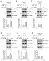
Involvement of multiple cysteine residues in DEM and H2O2 induced dimerization of STEP61. HEK293 cells expressing V5- and myc-tagged (A, D) STEP61 C65S/C76S, (B, E) STEP61 C472S or (C, F) STEP61 C65S/C76S/C472S were treated with (A–C) DEM for 6 hr or (D–F) H2O2 for 5 min. V5-tagged STEP61 was immunoprecipitated using anti-V5 antibody and co-immunoprecipitation of myc-tagged STEP61 was determined by probing the blots with anti-myc (upper panels) antibody. The blots were re-probed with and anti-V5 antibody (middle panels). Expression of myc-tagged STEP61 in total lysates was analyzed using anti-myc antibody (lower panels). Bar diagram represent mean ± SEM (n = 4–5) of co-immunoprecipitated myc-tagged STEP61. *p < 0.05.
Figure 8
Figure 8
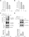
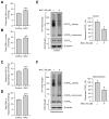
Effect of N-acetyl cysteine on GSH levels and STEP61 dimerization in cortex and hippocampus of aged rats. (A, B) Cortical and (C, D) hippocampal lysates from NAC treated rats were processed for quantification of both reduced and total GSH levels. Bar diagrams represent mean ± SEM of reduced and total GSH levels (n = 5). (E, F) In a parallel series of experiments cortical and hippocampal lysates treated with NAC were processed for immunoblot analysis under non-reducing conditions to assess STEP dimerization (upper panels). The same samples were processed under reducing conditions to assess STEP expression level (middle panels) and the blots were re-probed with anti-tubulin antibody (lower panels). Bar diagram represent mean ± SEM of dimerized STEP61 in non-reducing gels (n = 5). *p < 0.02, **p < 0.005; ***indicates p < 0.005.

Similar articles

Cited by

References

    1. Abe J, Okuda M, Huang Q, Yoshizumi M, Berk BC. Reactive oxygen species activate p90 ribosomal S6 kinase via Fyn and Ras. The Journal of biological chemistry. 2000;275:1739–1748. - PubMed
    1. Adam-Vizi V. Production of reactive oxygen species in brain mitochondria: contribution by electron transport chain and non-electron transport chain sources. Antioxidants & redox signaling. 2005;7:1140–1149. - PubMed
    1. Ballatori N, Krance SM, Notenboom S, Shi S, Tieu K, Hammond CL. Glutathione dysregulation and the etiology and progression of human diseases. Biological chemistry. 2009;390:191–214. - PMC - PubMed
    1. Boulanger LM, Lombroso PJ, Raghunathan A, During MJ, Wahle P, Naegele JR. Cellular and molecular characterization of a brain-enriched protein tyrosine phosphatase. J Neurosci. 1995;15:1532–1544. - PMC - PubMed
    1. Braak H, Braak E. Morphological criteria for the recognition of Alzheimer’s disease and the distribution pattern of cortical changes related to this disorder. Neurobiology of aging. 1994;15:355–356. discussion 379–380. - PubMed

Publication types

MeSH terms

LinkOut - more resources