Diagnostic imaging of psoriatic arthritis. Part I: etiopathogenesis, classifications and radiographic features
- PMID: 27104004
- PMCID: PMC4834372
- DOI: 10.15557/JoU.2016.0007
Diagnostic imaging of psoriatic arthritis. Part I: etiopathogenesis, classifications and radiographic features
Abstract
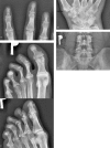
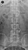
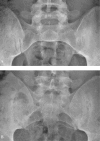
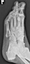
Psoriatic arthritis is one of the spondyloarthritis. It is a disease of clinical heterogenicity, which may affect peripheral joints, as well as axial spine, with presence of inflammatory lesions in soft tissue, in a form of dactylitis and enthesopathy. Plain radiography remains the basic imaging modality for PsA diagnosis, although early inflammatory changes affecting soft tissue and bone marrow cannot be detected with its use, or the image is indistinctive. Typical radiographic features of PsA occur in an advanced disease, mainly within the synovial joints, but also in fibrocartilaginous joints, such as sacroiliac joints, and additionally in entheses of tendons and ligaments. Moll and Wright classified PsA into 5 subtypes: asymmetric oligoarthritis, symmetric polyarthritis, arthritis mutilans, distal interphalangeal arthritis of the hands and feet and spinal column involvement. In this part of the paper we discuss radiographic features of the disease. The next one will address magnetic resonance imaging and ultrasonography.
Łuszczycowe zapalenie stawów jest jednostką należącą do grupy zapaleń kręgosłupa z towarzyszącym zapaleniem stawów obwodowych (spondyloarthritis, SpA). Choroba ma różne manifestacje kliniczne – może przebiegać z zajęciem stawów obwodowych, jak również kręgosłupa osiowego oraz z obecnością zmian zapalnych tkanek miękkich, w postaci zapalenia palca (dactylitis) bądź entezopatii. Klasyczna radiografia stanowi metodę z wyboru używaną w diagnostyce łuszczycowego zapalenia stawów, jednak nie uwidacznia ona wczesnych zmian zapalnych tkanek miękkich i szpiku kostnego lub obraz radiograficzny nie jest charakterystyczny. Typowe zmiany radiograficzne ujawniają się w zaawansowanych stadiach choroby; dotyczą głównie stawów maziówkowych, lecz także chrzęstno-włóknistych, takich jak stawy krzyżowo-biodrowe, oraz entez ścięgien i więzadeł. Moll i Wright sklasyfikowali łuszczycowe zapalenie stawów, wyróżniając pięć podtypów choroby: asymetryczne zapalenie nielicznostawowe, symetryczne zapalenie wielostawowe, postać nadżerkową arthritis mutilans, postać zajmującą stawy międzypaliczkowe dalsze palców rąk i stóp oraz zajęcie kręgosłupa osiowego. W pierwszej części artykułu przedstawiono radiograficzne manifestacje choroby. W części drugiej zostaną omówione zmiany widoczne w badaniu metodą rezonansu magnetycznego i w ultrasonografii.
Keywords: diagnostic imaging; enthesitis; plain radiography; psoriatic arthritis; spondyloarthritis.
Figures


Similar articles
-
Diagnostic imaging of psoriatic arthritis. Part II: magnetic resonance imaging and ultrasonography.J Ultrason. 2016 Jun;16(65):163-74. doi: 10.15557/JoU.2016.0018. Epub 2016 Jun 29. J Ultrason. 2016. PMID: 27446601 Free PMC article. Review.
-
Influence of Disease Manifestations on Health-related Quality of Life in Early Psoriatic Arthritis.J Rheumatol. 2018 Nov;45(11):1526-1531. doi: 10.3899/jrheum.171406. Epub 2018 Jul 1. J Rheumatol. 2018. PMID: 29961685
-
Clinical features of psoriatic arthritis.Best Pract Res Clin Rheumatol. 2021 Jun;35(2):101670. doi: 10.1016/j.berh.2021.101670. Epub 2021 Mar 17. Best Pract Res Clin Rheumatol. 2021. PMID: 33744078 Review.
-
The other arthritides. Roentgenologic features of osteoarthritis, erosive osteoarthritis, ankylosing spondylitis, psoriatic arthritis, Reiter's disease, multicentric reticulohistiocytosis, and progressive systemic sclerosis.Radiol Clin North Am. 1988 Nov;26(6):1195-212. Radiol Clin North Am. 1988. PMID: 3051093 Review.
-
Psoriatic arthritis: imaging techniques.Reumatismo. 2012 Jun 5;64(2):99-106. doi: 10.4081/reumatismo.2012.99. Reumatismo. 2012. PMID: 22690386 Review.
Cited by
-
The Involvement of Temporomandibular Joint in Psoriatic Arthritis: A Report of a Rare Case.Cureus. 2021 Dec 13;13(12):e20392. doi: 10.7759/cureus.20392. eCollection 2021 Dec. Cureus. 2021. PMID: 35036222 Free PMC article.
-
Hand Erosive Osteoarthritis and Distal Interphalangeal Involvement in Psoriatic Arthritis: The Place of Conservative Therapy.J Clin Med. 2021 Jun 15;10(12):2630. doi: 10.3390/jcm10122630. J Clin Med. 2021. PMID: 34203754 Free PMC article. Review.
-
Gout and 'Podagra' in medieval Cambridge, England.Int J Paleopathol. 2021 Jun;33:170-181. doi: 10.1016/j.ijpp.2021.04.007. Epub 2021 May 4. Int J Paleopathol. 2021. PMID: 33962231 Free PMC article.
-
Enthesitis and Dactylitis in Psoriatic Disease: A Guide for Dermatologists.Am J Clin Dermatol. 2018 Dec;19(6):839-852. doi: 10.1007/s40257-018-0377-2. Am J Clin Dermatol. 2018. PMID: 30117018 Free PMC article. Review.
-
Proteoglycan loss in the articular cartilage is associated with severity of joint inflammation in psoriatic arthritis-a compositional magnetic resonance imaging study.Arthritis Res Ther. 2020 May 29;22(1):124. doi: 10.1186/s13075-020-02219-7. Arthritis Res Ther. 2020. PMID: 32471515 Free PMC article.
References
-
- Rudwaleit M, Taylor WJ. Classification criteria for psoriatic arthritis and ankylosing spondylitis/axial spondyloarthritis. Best Prac Res Clin Rheumatol. 2010;24:589–604. - PubMed
-
- Wright V. Psoriatic arthritis. A comparative study of rheumatoid arthritis, psoriasis, and arthritis associated with psoriasis. AMA Arch Derm. 1959;80:27–35. - PubMed
-
- Paparo F, Ravelli M, Semprini A, Camellino D, Garlaschi A, Cimmino MA, et al. Seronegative spondyloarthropathies: what radiologists should know. Radiol Med. 2014;119:156–163. - PubMed
-
- Ory PA. Radiography in the assessment of musculoskeletal conditions. Best Pract Res Clin Rheumatol. 2003;17:495–512. - PubMed
-
- Sokolik R, Szechiński J. Łuszczycowe zapalenie stawów. In: Wiland P, editor. Reumatologia 2010/2011 – nowe trendy. Poznań: Termedia; 2011. pp. 99–114.
Publication types
LinkOut - more resources
Full Text Sources
Other Literature Sources
Research Materials
Miscellaneous
