

doi: 10.1055/s-0036-1582475.
Epub 2016 Apr 13.
In Vivo High-Resolution Trabecular Microstructure of Kienböck Lunate before and after Radial Shortening: A Case Report
Affiliations
- PMID: 27104074
- PMCID: PMC4838466
- DOI: 10.1055/s-0036-1582475
Item in Clipboard
In Vivo High-Resolution Trabecular Microstructure of Kienböck Lunate before and after Radial Shortening: A Case Report
J Wrist Surg.
2016 May.
Abstract
We report a patient with stage IIIB Kienböck disease treated with radial shortening where preoperative and sequential postoperative imaging were done using in vivo high-resolution peripheral quantitative micro-computed tomography (micro-CT) scan. Sequential in vivo micro-CT scan analysis of a target zone of the Kienböck lunate of this patient demonstrated early signs of lunate remodeling (bone trabecular densification) at 5-month follow-up suggesting an ongoing healing process. These early remodeling micro-CT scan signs were confirmed at 5 years' follow-up as well.
Keywords: HR-pQCT; Kienböck disease; lunate; quantitative micro-CT scan.
Conflict of interest statement
Figures
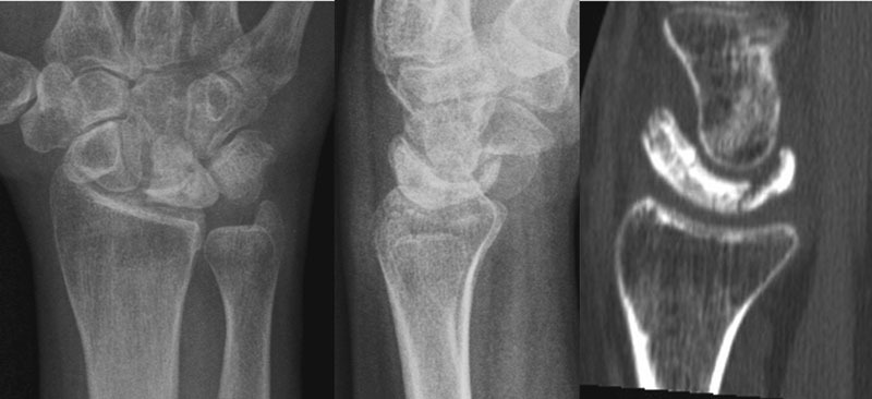
Preoperative PA and lateral standard radiographs and conventional CT scan (sagittal slice) of Kienböck right lunate of our patient.
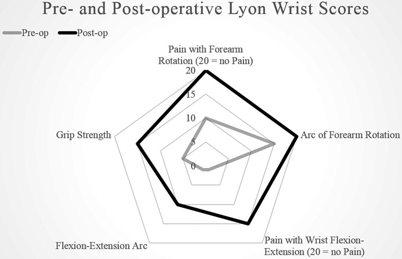
Diagrammatic patterns of Lyon pre- and postoperative (5 years follow up) wrist scores in our patient.
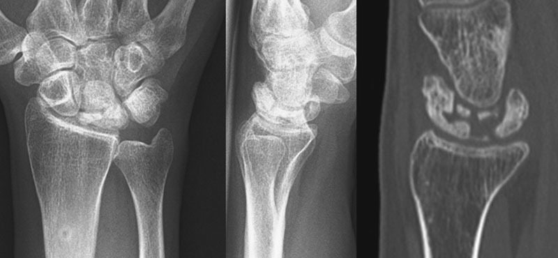
Postoperative 5 years' follow-up PA and lateral standard radiographs and conventional CT scan (sagittal slice) of Kienböck right lunate of our patient. Note the 2-mm radius shortening and the stabilization of the collapse.
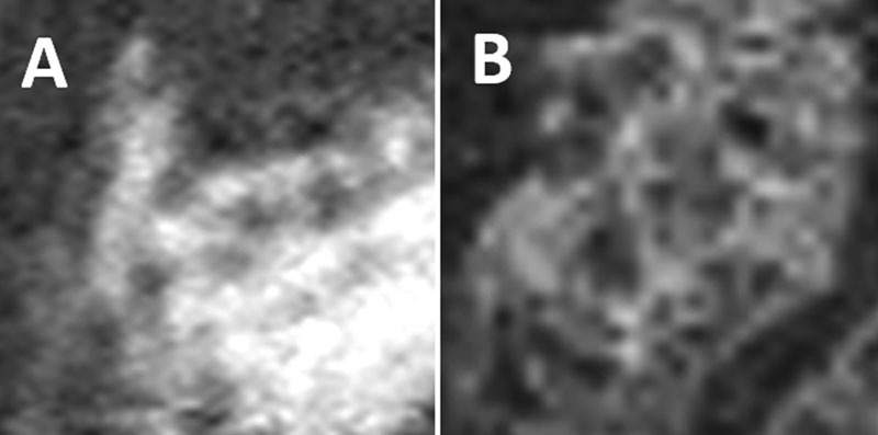
Trabecular microstructure as observed with HR-pQCT within Kienböck lunate VOI from preoperative (A) to 5 years' follow-up (B).
References
-
- Dias J J, Lunn P. Ten questions on Kienbock's disease of the lunate. J Hand Surg Eur Vol. 2010;35(7):538–543. - PubMed
-
- Irisarri C. Aetiology of Kienböck's disease. J Hand Surg [Br] 2004;29(3):281–287. - PubMed
-
- Owers K L, Scougall P, Dabirrahmani D, Wernecke G, Jhamb A, Walsh W R. Lunate trabecular structure: a radiographic cadaver study of risk factors for Kienbock's disease [corrected] J Hand Surg Eur Vol. 2010;35(2):120–124. - PubMed
-
- Makabe H, Iwasaki N, Kamishima T, Oizumi N, Tadano S, Minami A. Computed tomography osteoabsorptiometry alterations in stress distribution patterns through the wrist after radial shortening osteotomy for Kienböck disease. J Hand Surg Am. 2011;36(7):1158–1164. - PubMed
-
- Herzberg G, Mercier S, Charbonnier J P, Got P. Kienböck's disease in a 14-year-old gymnast: a case report. J Hand Surg Am. 2006;31(2):264–268. - PubMed
LinkOut - more resources
Full Text Sources
Other Literature Sources
