Radiotherapy in aggressive cutaneous pseudolymphoma: a case report and review of literature
- PMID: 27104170
- PMCID: PMC4831972
- DOI: 10.3857/roj.2016.34.1.76
Radiotherapy in aggressive cutaneous pseudolymphoma: a case report and review of literature
Abstract
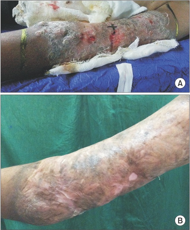
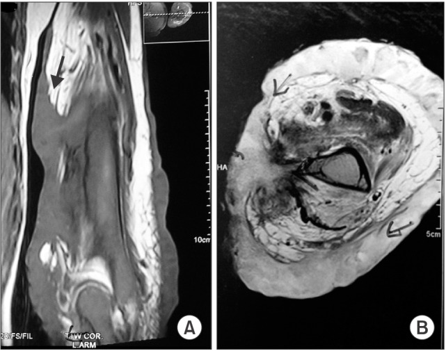
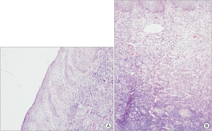
Pseudolymphoma is a nonspecific disease characterized by lesions with lymphomatous-appearing but benign accumulation of inflammatory cells. They generally present as small ulcero-nodular lesions confined to skin which often respond to local therapies. We describe an unusual presentation of an extensive and locally aggressive cutaneous pseudolymphoma in a 21-year-old male patient who presented with extensive cutaneous eruptions gradually progressing over 6 years to involve the entire circumference of his left arm. Magnetic resonance imaging scans of his left arm showed a lesion deeply infiltrating into the soft tissue reaching up to the humerus with intense periosteal reaction. He was successfully treated with radiotherapy after many failed attempts with surgery and chemotherapy.
Keywords: Pseudolymphoma; Radiotherapy.
Conflict of interest statement
Figures
Similar articles
-
Cutaneous pseudolymphoma: an unusual eyelid presentation.Semin Ophthalmol. 2012 Jan-Mar;27(1-2):35-6. doi: 10.3109/08820538.2011.622336. Semin Ophthalmol. 2012. PMID: 22352826
-
More advantages in detecting bone and soft tissue metastases from prostate cancer using 18F-PSMA PET/CT.Hell J Nucl Med. 2019 Jan-Apr;22(1):6-9. doi: 10.1967/s002449910952. Epub 2019 Mar 7. Hell J Nucl Med. 2019. PMID: 30843003
-
Cutaneous pseudolymphoma: an unusual presentation of a deep subcutaneous thigh mass.Ann Plast Surg. 1995 Nov;35(5):541-5. doi: 10.1097/00000637-199511000-00019. Ann Plast Surg. 1995. PMID: 8579278
-
Lymphoma or pseudolymphoma: A report of six cases and review of the literature.Dermatol Ther. 2019 Jul;32(4):e12807. doi: 10.1111/dth.12807. Epub 2019 Jan 7. Dermatol Ther. 2019. PMID: 30589489 Review.
-
HIV-related CD8+ cutaneous pseudolymphoma: efficacy of methotrexate.Dermatology. 2013;226(1):15-8. doi: 10.1159/000346242. Epub 2013 Jan 22. Dermatology. 2013. PMID: 23343593 Review.
Cited by
-
Upadacitinib in refractory cutaneous pseudolymphoma: A case report.SAGE Open Med Case Rep. 2025 Jun 19;13:2050313X251350370. doi: 10.1177/2050313X251350370. eCollection 2025. SAGE Open Med Case Rep. 2025. PMID: 40547414 Free PMC article.
References
-
- Ploysangam T, Breneman DL, Mutasim DF. Cutaneous pseudolymphomas. J Am Acad Dermatol. 1998;38(6 Pt 1):877–895. - PubMed
-
- Taylor RB, Fortney JA, Pollack RB, Metcalf JS, Jenrette JM. Radiation therapy for B-cell cutaneous lymphoid hyperplasia. Jpn J Radiol. 2010;28:385–387. - PubMed
-
- Safa G, Luce K, Darrieux L, Tisseau L, Ortonne N. Erythrodermic CD8+ pseudolymphoma during infliximab treatment in a patient with psoriasis: use of cyclosporine as a rescue therapy. J Am Acad Dermatol. 2014;71:e149–e150. - PubMed
-
- Kitagawa KH, Grassi M. Zoledronic acid-induced cutaneous B-cell pseudolymphoma. J Am Acad Dermatol. 2011;65:1238–1240. - PubMed
-
- Ingen-Housz-Oro S, Sbidian E, Ortonne N, et al. HIV-related CD8+ cutaneous pseudolymphoma: efficacy of methotrexate. Dermatology. 2013;226:15–18. - PubMed
LinkOut - more resources
Full Text Sources
Other Literature Sources