Review
. 2016 Sep 2;10(5):568-575.
doi: 10.1080/19336918.2016.1173803. Epub 2016 Apr 22.

Toward correlating structure and mechanics of platelets

Affiliations
Review

Toward correlating structure and mechanics of platelets

Simona Sorrentino et al. Cell Adh Migr. .

Abstract

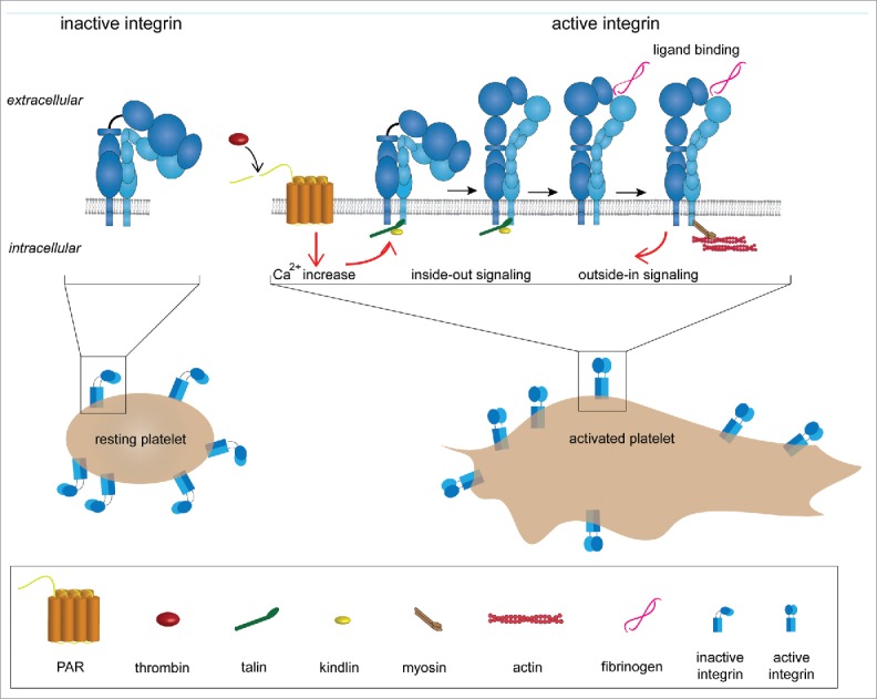
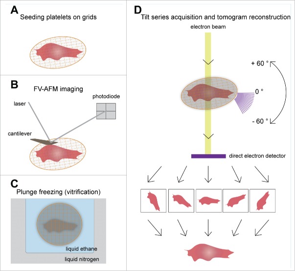
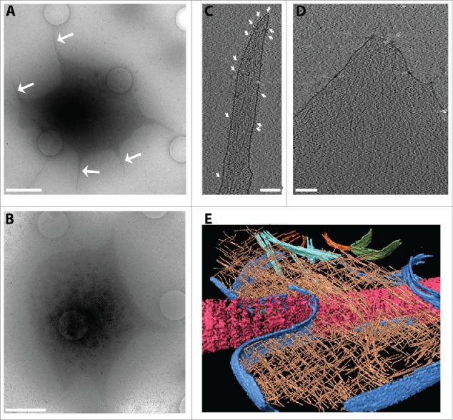
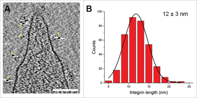
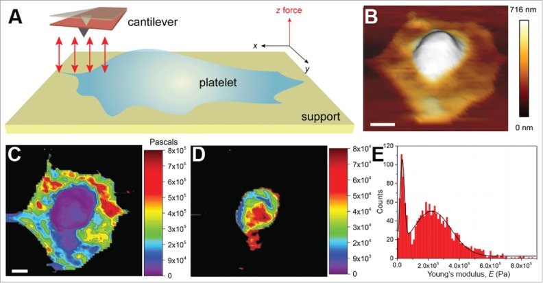
The primary physiological function of blood platelets is to seal vascular lesions after injury and form hemostatic thrombi in order to prevent blood loss. This task relies on the formation of strong cellular-extracellular matrix interactions in the subendothelial lesions. The cytoskeleton of a platelet is key to all of its functions: its ability to spread, adhere and contract. Despite the medical significance of platelets, there is still no high-resolution structural information of their cytoskeleton. Here, we discuss and present 3-dimensional (3D) structural analysis of intact platelets by using cryo-electron tomography (cryo-ET) and atomic force microscopy (AFM). Cryo-ET provides in situ structural analysis and AFM gives stiffness maps of the platelets. In the future, combining high-resolution structural and mechanical techniques will bring new understanding of how structural changes modulate platelet stiffness during activation and adhesion.

Keywords: actin; atomic force microscopy; cryo-electron tomography; integrins; platelets.


Figures

Figure 1.
Figure 1.
Adhesion system of platelets (platelet spreading and integrin activation). In resting platelets, αIIbβ3 integrins are in a bent and inactive conformation. Integrin activation can be triggered in 2 ways: (1) inside-out signaling or (2) outside-in signaling. In the first case integrin activation is initiated by platelet agonists such as thrombin. Thrombin activates the protease-activated GPCRs (G protein-coupled receptors), which leads to an increase in concentration of the cytosolic Ca2+. This triggers an intracellular signaling cascade that promotes talin binding to the cytoplasmic tail of the β2 chain. This event elicits integrin extension and activation. In the outside-in signaling, αIIbβ3 is activated by ligand binding (e.g., fibrinogen).
Figure 2.
Figure 2.
Schematic overview of AFM-cryo ET correlative approach. (A) Purified platelets are seeded on ECM protein (fibrinogen or collagen type I) coated grids in the presence of 1 mM Ca2+ and Mg2+ to allow spreading. (B) Mechanical properties of the platelets are then measured by FV-AFM imaging. (C) After AFM, the sample could be vitrified by plunge freezing in liquid N2 cooled liquid ethane, and inserted into the cryo-electron microscope. (D) A series of images of the area of interest are acquired by tilting the stage from −60° to +60° with a 2° increment. The tilt series images are then back projected to reconstruct the 3D volume of the original platelet.
Figure 3.
Figure 3.
Analysis of platelet ultrastructure with cryo-electron tomography. Platelets were seeded on fibrinogen-coated (50 µg/ml) silicon grids in the presence of 1 mM Ca2+ and Mg2+ to allow spreading. (A, B) Overview images of platelets (4800x) at 30 µm defocus. In (A) the white arrows point to the filopodia emanating from the platelet during spreading. (B) An adhering platelet with a lamellopodium. Scale bars: 2 µm. (C) An x-y slice through a tomogram of a platelet filopodium. White arrows indicate densities assigned to integrins embedded in a continuous plasma membrane. Actin filaments are seen enclosed by the plasma membrane. (D) An x-y slice through a tomogram of a platelet lamellipodium. (E) A rendered view of a platelet spread on collagen fiber. Collagen (pink), plasma membrane (blue), actin (brown), microtubules (cyan) and mitochondria outer membrane (green) were segmented and rendered using Amira software (FEI). The cell thickness in these regions is typically 70–150 nm. Tomograms were acquired on an FEI Titan Krios, 300 kV, equipped with a Gatan Quantum Energy Filter and a K2 Summit direct electron detection camera, at 0.34 nm/pixel at the specimen level. The tilt series was acquired from −60o to +60o in 2° increments, at 8 µm defocus. The cumulative electron dose was ~40 e- Å-2.
Figure 4.
Figure 4.
Analysis of platelet membrane receptors. (A) An x-y slice through a tomogram of a platelet, where integrin αIIbβ3 has been immunogold labeled. Yellow stars highlight the 6 nm gold nanoparticles, which were observed only in the proximity of the platelet membrane. Scale bar: 100 nm. (B) Histogram of integrin length distribution. The integrin lengths were measured in 5 tomograms (acquired as in Figs. 1C, D) for a total of 366 integrins. The black line is a Gaussian fit to the histogram.
Figure 5.
Figure 5.
Mechanical investigation of platelets. (A) Schematic illustrating the principle of mechanical characterization of platelets. A platelet is allowed to spread on a fibrinogen-coated glass surface in a physiological buffer (e.g., Tyrode's buffer containing 1 mM each of Ca2+ and Mg2+ ions, ± thrombin). Unlike the conventional AFM contact mode imaging, here F-D curves are recorded (red arrows) in the x-y plane. The F-D curves are used to extract the desired material properties. (B) A 3D topograph of a platelet spread on a fibrinogen-coated surface. The platelet shows a typical fried-egg shape, a high central region and flat periphery, indicative of an adhering platelet. (C) F-D curves were acquired by indenting the platelet with a force of 500 pN at an approach-retract velocity of 5 µm/s. The extension curves were then fit to the Hertz model to obtain the Young's moduli, contour-mapped on the platelet surface. The central region was observed to be softer than the peripheral region. The stiff glass background is shown in black. (D) A resolution of ~137 nm (64 × 64 pixel map of 8.74 × 8.74 µm2) was reasonable to obtain a contour map specifically of the central region. (E) A histogram of the modulus values, 32 kPa for the central region and ~224 kPa for the peripheral region. Scale bars (B, C): 1 µm. C and D are the same scale.

Similar articles

Cited by

References

    1. Savage B, Almus-Jacobs F, Ruggeri ZM. Specific synergy of multiple substrate-receptor interactions in platelet thrombus formation under flow. Cell 1998; 94:657-66; PMID:9741630; http://dx.doi.org/10.1016/S0092-8674(00)81607-4 - DOI - PubMed
    1. Winokur R, Hartwig JH. Mechanism of shape change in chilled human platelets. Blood 1995; 85:1796-804; PMID:7703486. - PubMed
    1. Hartwig JH. The platelet: form and function. Semin Hematol 2006; 43:S94-100; PMID:16427392; http://dx.doi.org/10.1053/j.seminhematol.2005.11.004 - DOI - PubMed
    1. Geiger B, Yamada KM. Molecular architecture and function of matrix adhesions. Cold Spring Harb Perspect Biol 2011; 3:pii: a005033; PMID:21441590; http://dx.doi.org/10.1101/cshperspect.a005033 - DOI - PMC - PubMed
    1. Ablooglu AJ, Kang J, Petrich BG, Ginsberg MH, Shattil SJ. Antithrombotic effects of targeting alpha IIb beta 3 signaling in platelets. Blood 2009; 113:3585-92; PMID:19005179; http://dx.doi.org/10.1182/blood-2008-09-180687 - DOI - PMC - PubMed

Publication types

LinkOut - more resources