. 2016 Apr 22;11(4):e0153806.
doi: 10.1371/journal.pone.0153806. eCollection 2016.

Alterations of Dermal Connective Tissue Collagen in Diabetes: Molecular Basis of Aged-Appearing Skin

Affiliations

Alterations of Dermal Connective Tissue Collagen in Diabetes: Molecular Basis of Aged-Appearing Skin

Angela J Argyropoulos et al. PLoS One. .

Abstract

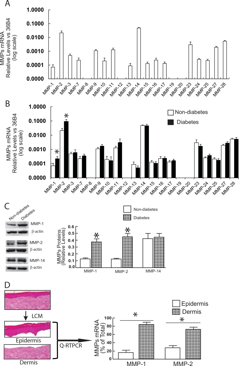
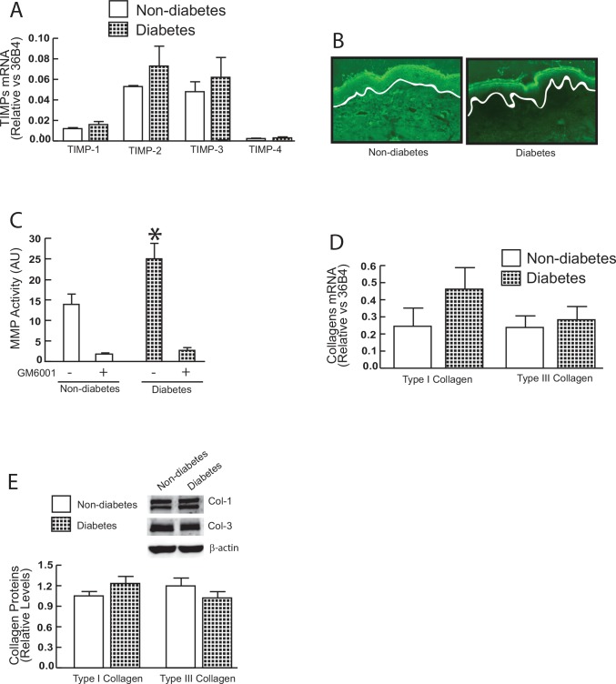
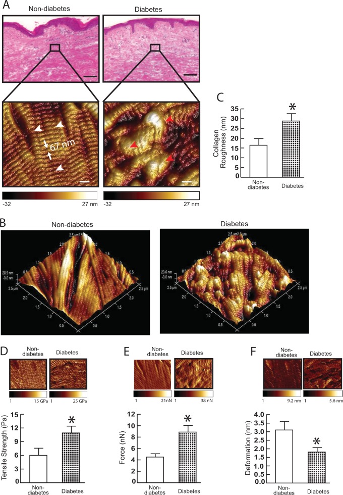
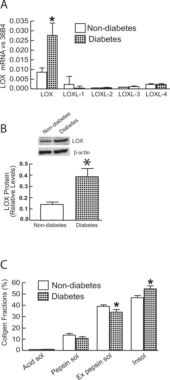
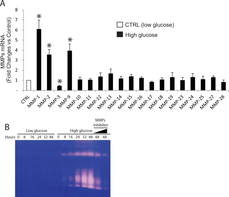
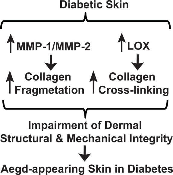
Alterations of the collagen, the major structural protein in skin, contribute significantly to human skin connective tissue aging. As aged-appearing skin is more common in diabetes, here we investigated the molecular basis of aged-appearing skin in diabetes. Among all known human matrix metalloproteinases (MMPs), diabetic skin shows elevated levels of MMP-1 and MMP-2. Laser capture microdissection (LCM) coupled real-time PCR indicated that elevated MMPs in diabetic skin were primarily expressed in the dermis. Furthermore, diabetic skin shows increased lysyl oxidase (LOX) expression and higher cross-linked collagens. Atomic force microscopy (AFM) further indicated that collagen fibrils were fragmented/disorganized, and key mechanical properties of traction force and tensile strength were increased in diabetic skin, compared to intact/well-organized collagen fibrils in non-diabetic skin. In in vitro tissue culture system, multiple MMPs including MMP-1 and MM-2 were induced by high glucose (25 mM) exposure to isolated primary human skin dermal fibroblasts, the major cells responsible for collagen homeostasis in skin. The elevation of MMPs and LOX over the years is thought to result in the accumulation of fragmented and cross-linked collagen, and thus impairs dermal collagen structural integrity and mechanical properties in diabetes. Our data partially explain why old-looking skin is more common in diabetic patients.


Conflict of interest statement

Competing Interests: The authors have declared that no competing interests exist.

Figures

Fig 1
Fig 1. Elevated expression of MMP-1 and MMP-2 in diabetic human skin dermis in vivo.
(A) MMP-1 and MMP-2 mRNA levels were elevated in diabetic skin relative to non-diabetic control skin. Total RNA was extracted from skin tissue and mRNA levels were quantified by real-time RT-PCR. MMPs mRNA levels were normalized to the housekeeping gene 36B4, as an internal control for quantification. Data are relative levels to 36B4 (mean±SEM). MMPs mRNA levels were expressed by log scale. N = 12, *p<0.05. (B) MMP-1 and MMP-2 protein levels were elevated in diabetic human skin. Protein levels were determined by Western analysis and normalized by β-actin (loading control). Insets (left panel) show representative Western blots. Data are expressed as mean±SEM, N = 6, *p<0.05. (C) Elevated MMPs in diabetic dermis. Epidermis and dermis were captured by Laser Capture Microdissection (LCM, see Methods for details). N = 6, *p<0.05. Total RNA was extracted from captured epidermis and dermis, and mRNA levels were quantified by real-time RT-PCR. MMPs mRNA levels were normalized to the housekeeping gene 36B4, as an internal control for quantification. Data are relative levels to 36B4 (mean±SEM). (D) MMP-1 and MMP-2, but not other MPs, were elevated in diabetic human skin dermis. Total RNA was extracted from the dermis captured by LCM and mRNA levels were quantified by real-time RT-PCR. MMPs mRNA levels were normalized to the housekeeping gene 36B4, as an internal control for quantification. Data are relative levels to 36B4 (mean±SEM). N = 6, *p<0.05.
Fig 2
Fig 2. MMP activity, TIMPs, and collagen expression in diabetic human skin in vivo.
(A) TIMP mRNA expression in diabetic and non-diabetic control skin. TIMP mRNA levels were normalized to the housekeeping gene 36B4, as an internal control for quantification. Data are relative levels to 36B4 (mean±SEM) N = 6. (B) Elevated MMP activity in the dermis of photodamaged forearm skin determined by in situ zymography. Loss of green fluorescence in diabetic dermis indicates degradation of fluorescein-collagen substrate. White lines indicate boundary between the epidermis (top) and dermis (bottom). N = 6. (C) MMP activity in diabetic and non-diabetic control skin (see Methods for details). N = 9–11. (D) Type I and type III procollagen mRNA expression in diabetic and non-diabetic control skin. Type I and type III procollagen mRNA levels were normalized to the housekeeping gene 36B4, as an internal control for quantification. Data are relative levels to 36B4 (mean±SEM) N = 6. (E) Type I and type III collagen protein levels were determined by Western analysis and normalized by β-actin (loading control). Insets (left panel) show representative Western blots. Data are expressed as mean±SEM, N = 6.
Fig 3
Fig 3. Collagen fibril nanoscale morphology and mechanical properties in diabetic skin.
(A) Nanoscale collagen fibrils were imaged by atomic force microscopy (AFM). The white and red arrow heads indicate intact and fragmented/disorganized collagen fibrils, respectively. Images are representative of nine subjects. (B) Three-dimensional nanoscale collagen fibrils were imaged by AFM. Images are representative of nine subjects. (C) Collagen fibril roughness was analyzed using Nanoscope Analysis software (Nanoscope_Analysis_v120R1sr3, Bruker-AXS, Santa Barbara, CA). Results are expressed as the mean ± SEM, N = 6, *p<0.05. (D) Tensile strength, (E) traction forces, and (F) deformation were determined by AFM PeakForceTM Quantitative NanoMechanics mode and Nanoscope Analysis software. Means±SEM. N = 8, *p<0.05.
Fig 4
Fig 4. Alteration of lysyl oxidase (LOX) expression diabetic skin.
(A) LOX family members mRNA expression in diabetic and non-diabetic control skin. Lox family mRNA levels were normalized to the housekeeping gene 36B4, as an internal control for quantification. Data are relative levels to 36B4. Mean±SEM. N = 6. (B) LOX protein levels were determined by Western analysis and normalized by β-actin (loading control). Insets (left panel) show representative Western blots. Data are expressed as mean±SEM, N = 6, *p<0.05. (C) Collagen solubility by pepsin digestion was determined by HPLC (see Methods for details). Mean±SEM. N = 12, *p<0.05.
Fig 5
Fig 5. The effect of high glucose culture on MMPs mRNA levels and proteolytic activity in primary human skin dermal fibroblasts.
Cell were cultured for 48 h in low (5mM) or high (25 mM) glucose media (see Methods for details). The effects of high glucose on the levels of MMPs mRNA expression were quantified by real-time RT-PCR. MMPs mRNA levels were normalized to the housekeeping gene 36B4, as an internal control for quantification. Primary human skin dermal fibroblasts do not express MMP-8 and MMP-20. Mean±SEM. N = 4. *p<0.05. Proteolytic activities from the conditioned media were examined by zymography, as described in “Methods”. Areas of protease activity will appear as clear bands against a dark blue background where the protease has digested the substrate. Data are representative of three experiments. MMPs inhibitor (GM60001) was used as specificity of MMPs-mediated proteolytic activity (last two lanes).
Fig 6
Fig 6. Proposed model for alterations of dermal collagen structural and mechanical properties contribute to old-looking skin in diabetes.
Diabetic dermis constitutively expresses elevated levels of MMP-1/MMP-2 and LOX, which result in increased collagen fragmentation, crosslinking, and consequently alterations of dermal structural and mechanical properties of the dermis. Constitutive elevation of MMPs and LOX over decades is thought to result in accumulation of fragmented and cross-linked collagen fragments, and thus contribute to aged-appearing skin in diabetes.

References

    1. Duff M, Demidova O, Blackburn S, Shubrook J. Cutaneous manifestations of diabetes mellitus. Clinical diabetes: a publication of the American Diabetes Association. 2015;33(1):40–8. 10.2337/diaclin.33.1.40 - DOI - PMC - PubMed
    1. Demirseren DD, Emre S, Akoglu G, Arpaci D, Arman A, Metin A, et al. Relationship between skin diseases and extracutaneous complications of diabetes mellitus: clinical analysis of 750 patients. Am J Clin Dermatol. 2014;15(1):65–70. 10.1007/s40257-013-0048-2 - DOI - PubMed
    1. Levy L, Zeichner JA. Dermatologic manifestation of diabetes. J Diabetes. 2012;4(1):68–76. 10.1111/j.1753-0407.2011.00151.x - DOI - PubMed
    1. Perez MI, Kohn SR. Cutaneous manifestations of diabetes mellitus. Journal of the American Academy of Dermatology. 1994;30(4):519–31; quiz 32–4. . - PubMed
    1. Reiber GE. The epidemiology of diabetic foot problems. Diabet Med. 1996;13 Suppl 1:S6–11. . - PubMed

Publication types