. 2016 Apr 22;10(4):e0004601.
doi: 10.1371/journal.pntd.0004601. eCollection 2016 Apr.

Fasciola hepatica Surface Coat Glycoproteins Contain Mannosylated and Phosphorylated N-glycans and Exhibit Immune Modulatory Properties Independent of the Mannose Receptor

Affiliations

Fasciola hepatica Surface Coat Glycoproteins Contain Mannosylated and Phosphorylated N-glycans and Exhibit Immune Modulatory Properties Independent of the Mannose Receptor

Alessandra Ravidà et al. PLoS Negl Trop Dis. .

Abstract

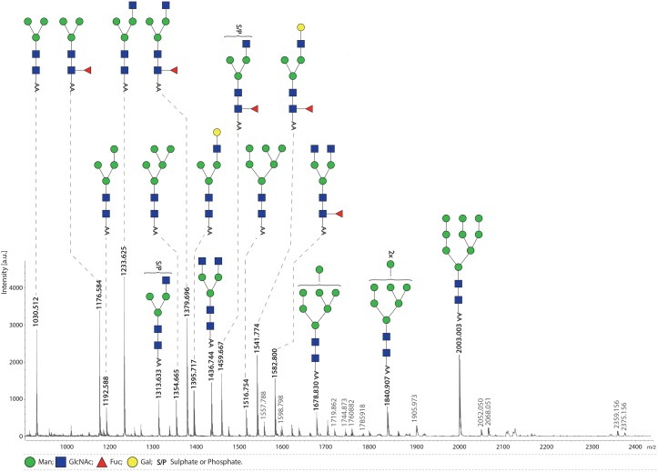
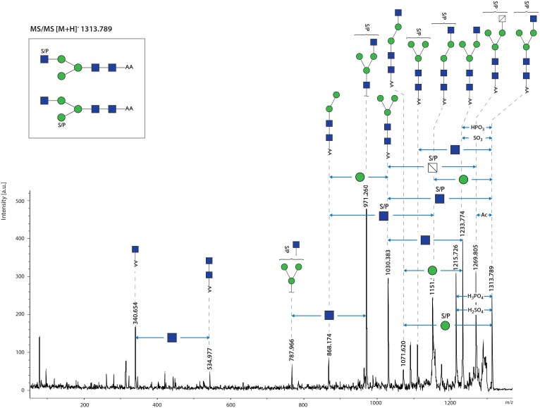
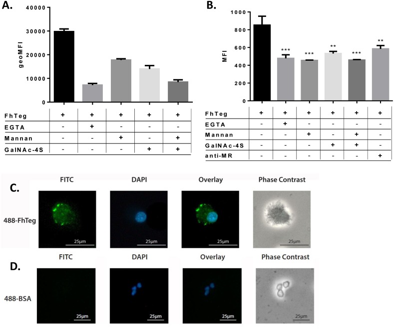
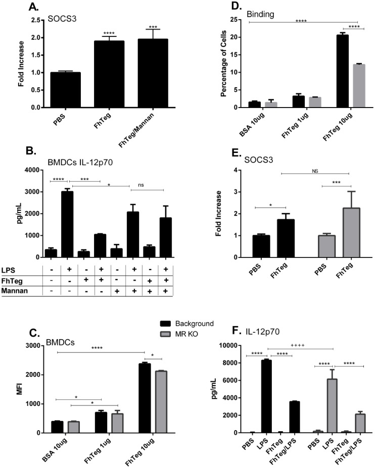
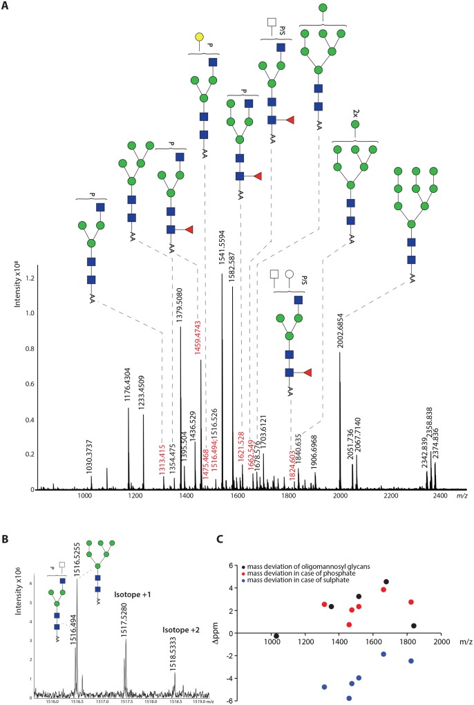
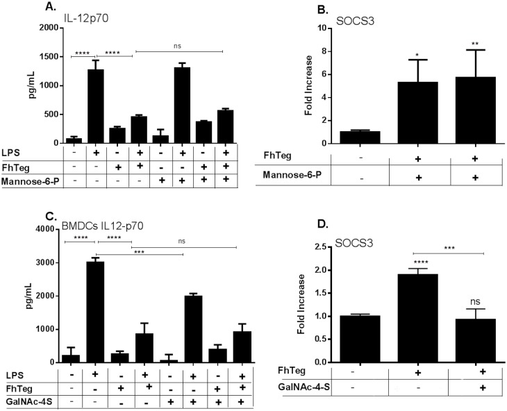
Fascioliasis, caused by the liver fluke Fasciola hepatica, is a neglected tropical disease infecting over 1 million individuals annually with 17 million people at risk of infection. Like other helminths, F. hepatica employs mechanisms of immune suppression in order to evade its host immune system. In this study the N-glycosylation of F. hepatica's tegumental coat (FhTeg) and its carbohydrate-dependent interactions with bone marrow derived dendritic cells (BMDCs) were investigated. Mass spectrometric analysis demonstrated that FhTeg N-glycans comprised mainly of oligomannose and to a lesser extent truncated and complex type glycans, including a phosphorylated subset. The interaction of FhTeg with the mannose receptor (MR) was investigated. Binding of FhTeg to MR-transfected CHO cells and BMDCs was blocked when pre-incubated with mannan. We further elucidated the role played by MR in the immunomodulatory mechanism of FhTeg and demonstrated that while FhTeg's binding was significantly reduced in BMDCs generated from MR knockout mice, the absence of MR did not alter FhTeg's ability to induce SOCS3 or suppress cytokine secretion from LPS activated BMDCs. A panel of negatively charged monosaccharides (i.e. GlcNAc-4P, Man-6P and GalNAc-4S) were used in an attempt to inhibit the immunoregulatory properties of phosphorylated oligosaccharides. Notably, GalNAc-4S, a known inhibitor of the Cys-domain of MR, efficiently suppressed FhTeg binding to BMDCs and inhibited the expression of suppressor of cytokine signalling (SOCS) 3, a negative regulator the TLR and STAT3 pathway. We conclude that F. hepatica contains high levels of mannose residues and phosphorylated glycoproteins that are crucial in modulating its host's immune system, however the role played by MR appears to be limited to the initial binding event suggesting that other C-type lectin receptors are involved in the immunomodulatory mechanism of FhTeg.


Conflict of interest statement

The authors have declared that no competing interests exist.

Figures

Fig 1
Fig 1. FhTeg preparation is rich in oligomannose and truncated complex type N-glycans carrying fucose or sulfate/phosphate moieties.
Fh tegumental antigens were digested with trypsin followed by PNGase F treatment. Released N-glycans were subsequently labelled with 2-AA and analysed by MALDI-TOF-MS in the negative ion-reflector mode. Signals are labelled with monoisotopic masses. Most abundant N-glycan structures are annotated in the spectrum while minor peaks are reported in the supplementing material (S1 Table). The signal at m/z 1582.8 [M-H]- is annotated according to MALDI-TOF/TOF-MS analysis.
Fig 2
Fig 2. FhTeg N-glycans carry core fucose and are sulphated/phosphorylated on terminal N-acetylglucosamine and mannose residues.
The sulphated/phosphorylated glycan observed at m/z 1313.6 [M-H]- in Fig 1 was analysed by MALDI-TOF- MS/MS. Differences in monosaccharides and functional groups content are indicated by double headed arrows.
Fig 3
Fig 3. Mannose-rich oligosaccharides are predominantly localized on scales and suckers of adult flukes.
High mannose type N-glycans are highly expressed on FhTeg as visualised in ConA fluorescence micrographs (A-D) and ConA blot (E) of adult Fasciola hepatica tegumental coat. A-D: Oral (OS) and Ventral (VS) suckers (A and B, respectively), tegumental spines (C-D) probed with fluorescein-labelled ConA (green); Scale bars: 200μm (A-B), 100μm (C), 50μm (D); spinelets details highlighted with arrows. E: Tegumental proteins (15μg) probed with biotin-labelled ConA/IR-labelled streptavidin complex (green), MW marker (Red).
Fig 4
Fig 4. FhTeg binding to dendritic cells is mediated by MR and is carbohydrate and calcium dependent.
A-B: MR-transfected CHO cells (A) and BMDCs (B) were stimulated with and without inhibitors, i.e. EGTA (10mM), anti-MR (1 μg ml-1), mannan (A: 100 μg ml-1;B: 1 mg/mL), GalNAc-4S (A: 1mM; B: 25 mM), for 45 min prior to stimulation with fluorescently labelled FhTeg (A: 1–10 μg ml-1; B: 5 μg/mL) for 45 min. Fluorescently labelled BSA was also used as control. FhTeg binding to cells was assessed by flow cytometry and reported in bar chart format. Data shown is the mean ± SD of one representative experiment; the experiment was repeated 2–3 times, **, p ≤ 0.01; ***, p ≤ 0.001 compared to FhTeg. C-D: BMDCs were stimulated with fluorescently labelled FhTeg (10μg ml-1, green) or BSA (10g ml-1, green)) for 45 min prior to paraformaldehyde fixation and mounting with DAPI (blue); Scale bar: 25μm.
Fig 5
Fig 5. The immune properties of FhTeg are independent of the MR receptor.
A: BMDCs were stimulated with mannan for 30 min prior to incubation with FhTeg for 2.5 h. Total RNA was extracted, and after reverse transcription cDNA was analyzed with qPCR for SOCS3. RNA expression was normalized to GAPDH and actin control genes. B: BMDCs were pre-incubated with mannan prior stimulation with PBS or FhTeg (10μg) before addition of LPS (100ng ml-1) for 18 h. IL12p70 levels were measured with commercial ELISA kits. Data are presented as the mean ± SEM of two independent experiments. ***p ≤ 0.001; ****p ≤ 0.0001 compared to LPS group. C,D: BMDCs isolated from MR-knockout mice were stimulated with fluorescently labelled FhTeg or BSA (10μg ml-1, green) for 45 min prior to paraformaldehyde fixation. FhTeg binding to cells was assessed by flow cytometry and reported in bar chart format. E: BMDCs isolated from MR-knockout mice were stimulated with FhTeg (10μg) for 2.5 h. Total RNA was extracted, and after reverse transcription cDNA was analyzed with qPCR for SOCS3. RNA expression was normalized to GAPDH and actin control genes. F. BMDCs derived from MR knockout mice were stimulated with PBS or FhTeg (10μg) before addition of LPS (100ng ml-1) for 18 h. IL12p70 levels were measured with commercial ELISA kits. Data are presented as the mean ± SEM of two independent experiments. *p ≤ 0.05, **, p ≤ 0.01; ***, p ≤ 0.001.
Fig 6
Fig 6. MALDI-FT-ICR-MS analysis of FhTeg N-glycans indicates the presence of phosphorylated glycan species.
A: FT-ICR-MS spectrum indicating the phosphorylated glycans identified by accurate mass determination with external calibration and additional comparison with the internal confirmed oligomannose glycans. B: Zoom region showing the high resolution separation between the Man6GlcNAc2 glycan and a phosphorylated glycan of almost identical mass. C: The scatter plot indicates the deviation of the measured m/z from the calculated m/z of phosphorylated glycans in comparison with hypothetical sulphated glycans and the oligomannosyl glycans also present in the spectrum.
Fig 7
Fig 7. FhTeg–induced upregulation of SOCS3 transcription levels is reversed by anionic monosaccharide inhibitor GalNAc-4S and not Man-6P.
A,C: BMDCs were pre-incubated with Man-6P (A) or GalNAc-4S (C) prior stimulation with PBS or FhTeg (10μg) before addition of LPS (100ng ml-1) for 18 h. IL12p70 levels were measured with commercial ELISA kits. Data are presented as the mean ± SEM of two independent experiments. ***p ≤ 0.001; ****p ≤ 0.0001 compared to LPS group. B,D: BMDCs were stimulated with Man-6P (B) or GalNAc-4S (D) for 30 min prior to incubation with FhTeg for 2.5 h. Total RNA was extracted, and after reverse transcription cDNA was analyzed with qPCR for SOCS3. RNA expression was normalized to GAPDH and actin control genes. C: BMDCs were stimulated with GalNAc-4S for 30 min prior to incubation with FhTeg for 2.5 h. Total RNA was extracted, and after reverse transcription cDNA was analyzed with qPCR for SOCS3. RNA expression was normalized to GAPDH and actin control genes.

References

    1. Allen JE, Maizels RM. Diversity and dialogue in immunity to helminths. Nat Rev Immunol. 2011;11: 375–388. 10.1038/nri2992 - DOI - PubMed
    1. McSorley HJ, Hewitson JP, Maizels RM. Immunomodulation by helminth parasites: defining mechanisms and mediators. Int J Parasitol. 2013; 43:301–310. 10.1016/j.ijpara.2012.11.011 - DOI - PubMed
    1. Taylor MD, van der Werf N, Maizels RM. T cells in helminth infection: the regulators and the regulated. Trends Immunol. 2012; 33:181–189. 10.1016/j.it.2012.01.001 - DOI - PubMed
    1. Vásquez-Mendoza A, Carrero JC, Rodriguez-Sosa M. Parasitic infections: a role for C-type lectin receptors. Biomed Res Int. 2013; 456352. - PMC - PubMed
    1. Meevissen MH, Yazdanbakhsh M, Hokke CH. Schistosoma mansoni egg glycoproteins and C-type lectins of host immune cells: molecular partners that shape immune responses. Exp Parasitol. 2012; 132: 14–21. 10.1016/j.exppara.2011.05.005 - DOI - PubMed

Publication types

MeSH terms

LinkOut - more resources