Review
. 2016 Apr 21;165(3):524-34.
doi: 10.1016/j.cell.2016.03.047.

Targeting Neural Circuits

Affiliations
Review

Targeting Neural Circuits

Priyamvada Rajasethupathy et al. Cell. .

Abstract

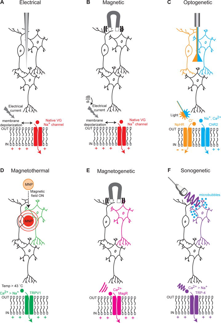
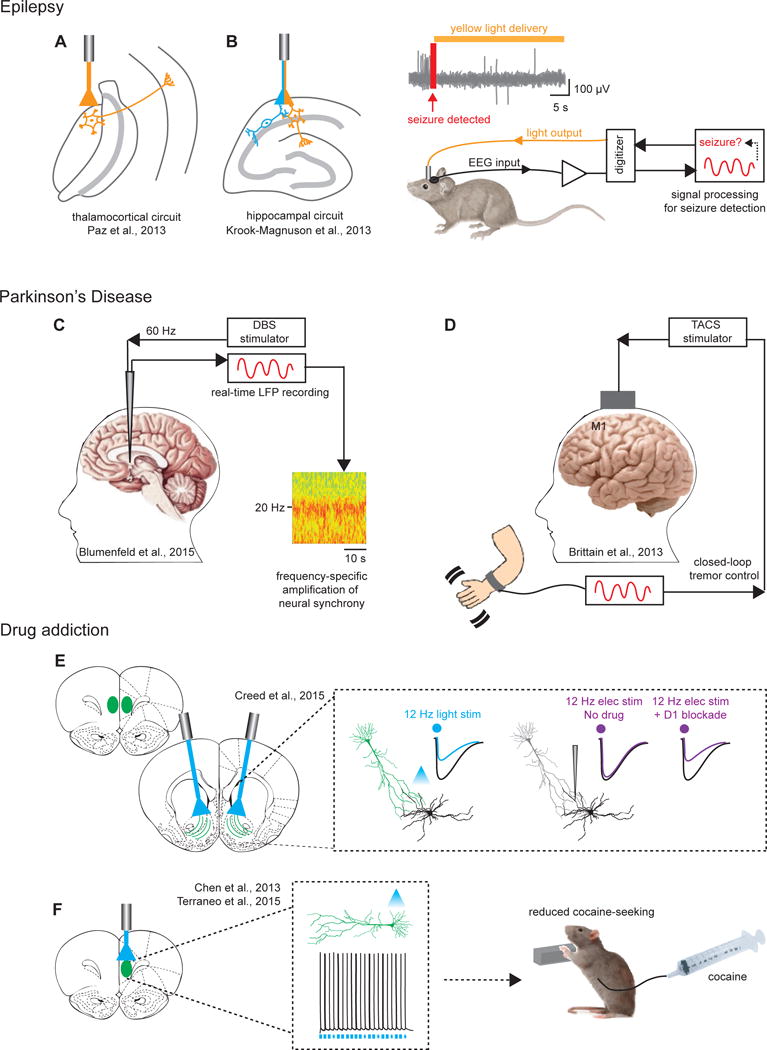
Optogenetic methodology enables direct targeting of specific neural circuit elements for inhibition or excitation while spanning timescales from the acute (milliseconds) to the chronic (many days or more). Although the impact of this temporal versatility and cellular specificity has been greater for basic science than clinical research, it is natural to ask whether the dynamic patterns of neural circuit activity discovered to be causal in adaptive or maladaptive behaviors could become targets for treatment of neuropsychiatric diseases. Here, we consider the landscape of ideas related to therapeutic targeting of circuit dynamics. Specifically, we highlight optical, ultrasonic, and magnetic concepts for the targeted control of neural activity, preclinical/clinical discovery opportunities, and recently reported optogenetically guided clinical outcomes.


Figures

Figure 1
Figure 1. Technologies for targeting specified regions & circuits
(A) Extracellular electrical stimulation of neurons through application of current; with regional targeting capability but without neuron-type specificity, capacitance currents are driven that lead to membrane depolarization, opening of native voltage-gated sodium channels, and further depolarization of the membrane with spike firing. (B) Stimulation of neurons through application of a transcranial magnetic field; again with regional targeting capability but without cell-type specificity, rapidly-changing magnetic fields (dB/dt) induce electrical currents in tissue and spike firing as in (A). (C) Cell type-specific optical stimulation or inhibition of neurons expressing light-sensitive excitatory cation channels (for example the channelrhodopsin ChR2) or inhibitory pumps (for example the halorhodopsin NpHR) that are expressed in a cell-type specific manner (here depicted by NpHR expressed specifically in the orange cell and ChR2 specifically in the blue cell). (D) Magnetothermal activation of neurons through application of a magnetic field targeted toward magnetic nanoparticles (MNP) that can transduce magnetic into thermal energy capable of opening heat sensitive (TRPV1) depolarizing channels, which can be expressed in a cell-type specific manner (here depicted as the green neuron). Generalizable AAV-based cell-type targeting strategies as developed for optogenetics may also be used to target TRP channels. (E) Application of a magnetic field that can open magnetically-sensitive channels (MagR) allowing the influx of Ca2+ with the potential for cell cell type-specific activation (here depicted as the pink neuron). (F) Ultrasonic activation of neurons through application of low pressure ultrasound waves that are transduced by gas-filled microbubbles into mechanical energy sufficient to open mechanosensitive (TRP-4) channels, which might also be expressed in a cell-type specific manner (here depicted as the purple neuron).
Figure 2
Figure 2. Disease -related circuit-targeting demonstrations
(A) Targeting thalamocortical neurons and (B) hippocampal neurons with closed-loop strategies to cause real-time interruption of EEG- and behaviorally-defined seizures. Right panel in (B): Depiction of closed-loop setup wherein EEG inputs are used to detect seizure onset, following which real-time inhibition can be administered through optogenetic manipulation. (C) Low frequency DBS stimulation (60Hz), in contrast to traditional high frequency stimulation (130 Hz), results in significant amplification of subthalamic neural synchrony and alpha/beta band power thought to underlie improved gait symptoms in Parkinson’s. (D) Use of an alternative non-invasive approach termed transcranial alternating current stimulation (TACS) is depicted for stimulation of motor cortex; guided by real-time readout of cortical oscillations linked to tremor, TACS was found to result in significant cancellation of the resting state tremor. (E) 12 Hz optical stimulation was reported in this study to produce robust LTD, whereas 12 Hz electrical stimulation did not. The discrepancy for these investigators was attributed to off-target recruitment of D1 receptors by electrical stimulation which impeded LTD; a combination of low frequency DBS together with D1 antagonists provided reliable LTD and restoration of behavior in cocaine seeking animals. (F) Optogenetic prelimbic cortical excitation significantly prevented cocaine-seeking in rats (by compensating for hypoactivity observed in cocaine-seeking rats), pointing to the prefrontal cortex as a promising therapeutic target for compulsive drug use.

References

    1. Akerboom J, Carreras Calderón N, Tian L, Wabnig S, Prigge M, Tolö J, Gordus A, Orger MB, Severi KE, Macklin JJ, et al. Genetically encoded calcium indicators for multi-color neural activity imaging and combination with optogenetics. Front Mol Neurosci. 2013;6:2. - PMC - PubMed
    1. Adamantidis A, Zhang F, Aravanis A, Deisseroth K, de Lecea L. Neural substrates of awakening probed with optogenetic control of hypocretin neurons. Nature. 2007;450:420–4. - PMC - PubMed
    1. Aravanis A, Wang LP, Zhang F, Meltzer L, Mogri M, Schneider MB, Deisseroth K. An optical neural interface. Journal of Neural Engineering. 2007;4:S143–S156. - PubMed
    1. Berndt A, Yizhar O, Gunaydin LA, Hegemann P, Deisseroth K. Bi-stable neural state switches. Nature Neuroscience. 2008;12:229–34. - PubMed
    1. Berndt A, Lee SY, Ramakrishnan C, Deisseroth K. Structure-guided transformation of channelrhodopsin into a light-activated chloride channel. Science. 2014;344:420–424. - PMC - PubMed