

Functional independence in resting-state connectivity facilitates higher-order cognition
- PMID: 27105037
- PMCID: PMC5233432
- DOI: 10.1016/j.bandc.2016.03.008
Functional independence in resting-state connectivity facilitates higher-order cognition
Abstract
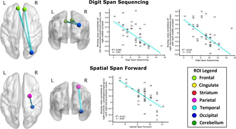
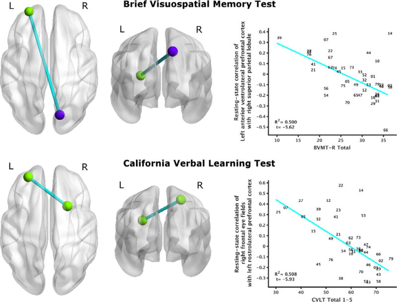
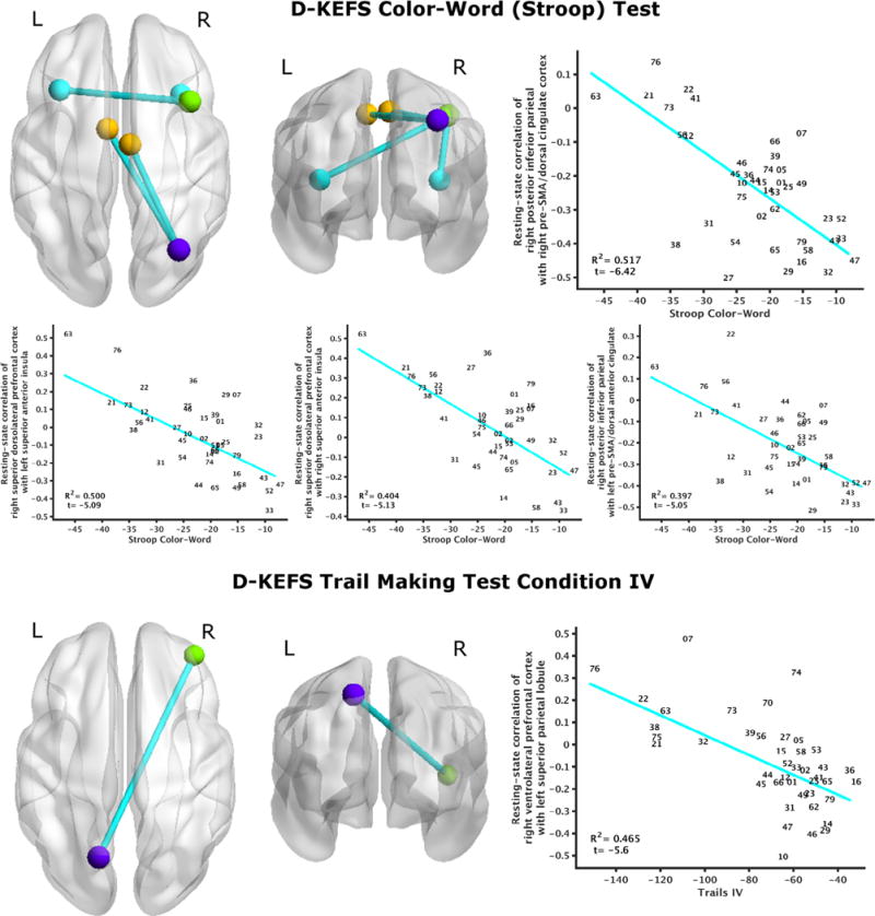
Growing evidence suggests that intrinsic functional connectivity (i.e. highly structured patterns of communication between brain regions during wakeful rest) may encode cognitive ability. However, the generalizability of these findings is limited by between-study differences in statistical methodology and cognitive domains evaluated. To address this barrier, we evaluated resting-state neural representations of multiple cognitive domains within a relatively large normative adult sample. Forty-four participants (mean(sd) age=31(10) years; 18 male and 26 female) completed a resting-state functional MRI scan and neuropsychological assessments spanning motor, visuospatial, language, learning, memory, attention, working memory, and executive function performance. Robust linear regression related cognitive performance to resting-state connectivity among 200 a priori determined functional regions of interest (ROIs). Only higher-order cognitions (such as learning and executive function) demonstrated significant relationships between brain function and behavior. Additionally, all significant relationships were negative - characterized by moderately positive correlations among low performers and weak to moderately negative correlations among high performers. These findings suggest that functional independence among brain regions at rest facilitates cognitive performance. Our interpretation is consistent with graph theoretic analyses which represent the brain as independent functional nodes that undergo dynamic reorganization with task demand. Future work will build upon these findings by evaluating domain-specific variance in resting-state neural representations of cognitive impairment among patient populations.
Keywords: Attention; Connectome; Executive functioning; Individual differences; Memory; fMRI.
Copyright © 2016 Elsevier Inc. All rights reserved.
Figures
References
-
- Alavash M, Doebler P, Holling H, Thiel CM, Giessing C. Is functional integration of resting state brain networks an unspecific biomarker for working memory performance? Neuroimage. 2015;108:182–193. - PubMed
-
- Albuquerque L, Loureiro C, Martins IP. Effect of lesion site on serial position during list learning: A study with the CVLT. International Journal of Neuroscience. 2008;118:917–933. - PubMed
-
- Alexander MP, Stuss DT, Fansabedian N. California verbal learning test: Performance by patients with focal frontal and non-frontal lesions. Brain. 2003;126:1493–1503. - PubMed
MeSH terms
Grants and funding
LinkOut - more resources
Full Text Sources
Other Literature Sources
