. 2016 Apr 22;12(4):e1005592.
doi: 10.1371/journal.ppat.1005592. eCollection 2016 Apr.

Legionella pneumophila-Derived Outer Membrane Vesicles Promote Bacterial Replication in Macrophages

Affiliations

Legionella pneumophila-Derived Outer Membrane Vesicles Promote Bacterial Replication in Macrophages

Anna Lena Jung et al. PLoS Pathog. .

Abstract

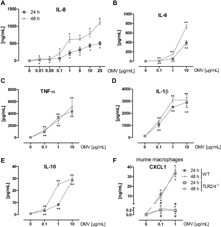
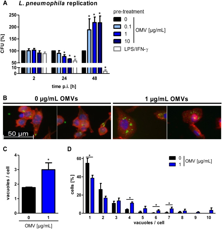
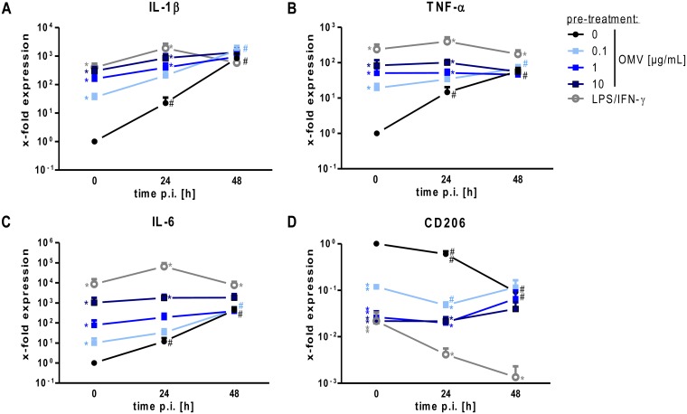
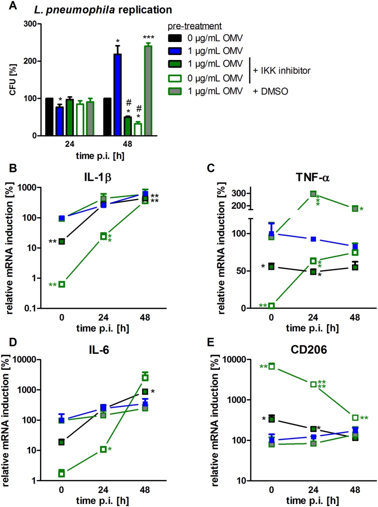
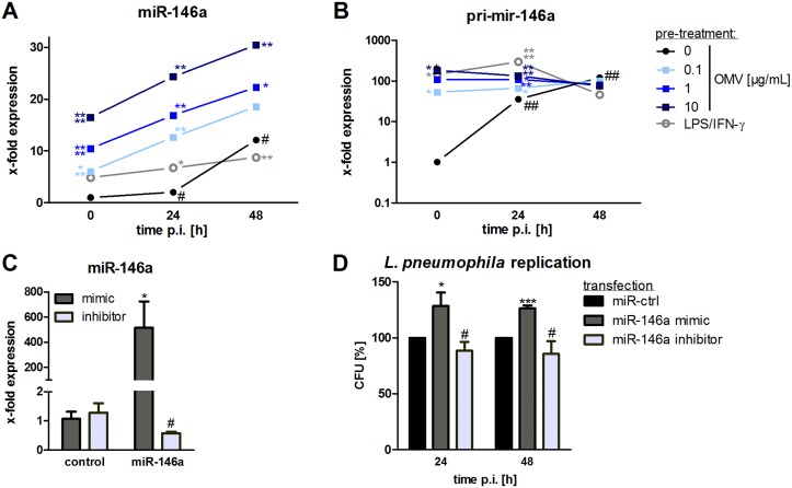
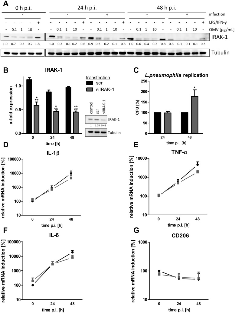
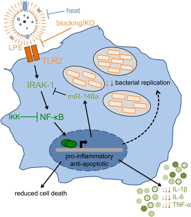
The formation and release of outer membrane vesicles (OMVs) is a phenomenon of Gram-negative bacteria. This includes Legionella pneumophila (L. pneumophila), a causative agent of severe pneumonia. Upon its transmission into the lung, L. pneumophila primarily infects and replicates within macrophages. Here, we analyzed the influence of L. pneumophila OMVs on macrophages. To this end, differentiated THP-1 cells were incubated with increasing doses of Legionella OMVs, leading to a TLR2-dependent classical activation of macrophages with the release of pro-inflammatory cytokines. Inhibition of TLR2 and NF-κB signaling reduced the induction of pro-inflammatory cytokines. Furthermore, treatment of THP-1 cells with OMVs prior to infection reduced replication of L. pneumophila in THP-1 cells. Blocking of TLR2 activation or heat denaturation of OMVs restored bacterial replication in the first 24 h of infection. With prolonged infection-time, OMV pre-treated macrophages became more permissive for bacterial replication than untreated cells and showed increased numbers of Legionella-containing vacuoles and reduced pro-inflammatory cytokine induction. Additionally, miRNA-146a was found to be transcriptionally induced by OMVs and to facilitate bacterial replication. Accordingly, IRAK-1, one of miRNA-146a's targets, showed prolonged activation-dependent degradation, which rendered THP-1 cells more permissive for Legionella replication. In conclusion, L. pneumophila OMVs are initially potent pro-inflammatory stimulators of macrophages, acting via TLR2, IRAK-1, and NF-κB, while at later time points, OMVs facilitate L. pneumophila replication by miR-146a-dependent IRAK-1 suppression. OMVs might thereby promote spreading of L. pneumophila in the host.


Conflict of interest statement

The authors have declared that no competing interests exist.

Figures

Fig 1
Fig 1. L. pneumophila OMVs induce cytokine secretion in macrophages.
(A) THP-1 cells were incubated with rising amounts of OMVs (0.01, 0.05, 0.1, 1, 5, 10, 25 μg/mL) for 24 or 48 h, respectively, or left untreated for control. Supernatant was collected for IL-8 ELISA. (B-E) THP-1 cells were incubated with rising amounts of OMVs (0.1, 1, 10 μg/mL) for 24 or 48 h, respectively, or left untreated for control. Supernatant was collected for magnetic multiplex ELISA. Results for IL-6 (B), TNF-α (C), IL-1β (D), and IL-10 (E) are shown. (F) mBMDM (wildtype (WT) and TLR2/4-/-) were stimulated with OMVs (0.1, 1 μg/mL) for 24 or 48 h, respectively, or left untreated for control. Supernatant was collected for CXCL1 ELISA. Mean values of three independent experiments are shown. Statistics: Mann-Whitney test; *p<0.05 and **p<0.01 compared to corresponding control. #p<0.05 compared to equally treated WT sample.
Fig 2
Fig 2. L. pneumophila OMVs alter bacterial replication and increase the vacuole amount per cell.
(A) THP-1 cells were pre-incubated for 20 h with different doses of OMVs (0.1, 1, 10 μg/mL) and were then infected with L. pneumophila (multiplicity of infection (MOI) of 0.5). Pre-stimulation with LPS (200 ng/mL) in combination with IFN-γ (200 ng/mL) served as a positive control for classically activated macrophages (M1). Bacterial replication after OMV pre-stimulation was determined by CFU assay. Cells were lysed after the indicated time points. Results are depicted relative to CFU count of not pre-treated (but infected) cells at every time point. (B) THP-1 cells were pre-treated with OMVs (1 μg/mL) for 20 h or left untreated before infection with L. pneumophila (MOI 0.5) for 48 h. Cells were fixed and stained with an α-LPS antibody (green), phalloidin (red), and DAPI (blue). One representative out of three independent experiments is shown. (C) Quantification of the number of vacuoles per cells of more than 70 cells per condition. Data are shown as mean values of three independent experiments. (D) Quantification of the distribution of vacuoles per cells. Mean values of three independent experiments are shown. Statistics: Mann-Whitney test; *p<0.05 compared to 0 μg/mL OMVs.
Fig 3
Fig 3. OMVs reduce responsiveness of macrophages to L. pneumophila infection.
(A-D) THP-1 cells treated as described in Fig 2A and RNA samples were taken at the time of infection (0 h) or 24 and 48 h post infection (p.i.). qPCR was performed for markers of classically activated macrophages (A: IL-1β, B: TNF-α, C: IL-6) and alternatively activated macrophages (D: CD206). Results are normalized to untreated control cells at every time point. Mean values of three independent experiments are shown. Statistics: Mann-Whitney test; * p<0.05 compared to 0 μg/mL OMVs. # p<0.05 compared to equally treated 0 h sample.
Fig 4
Fig 4. Blocking of NF-κB signaling prior to OMV treatment reduces bacterial replication and M1 activation.
THP-1 cells were pre-incubated with IKK inhibitor (1 μM) 90 min before OMV stimulation (1 μg/mL). DMSO was used as a solvent control. After 20 h of OMV incubation, cells were infected with L. pneumophila (MOI 0.5) and lysed for CFU 24 and 48 h p.i.. Results are depicted relative to CFU count of not pre-treated (but infected) cells (A). RNA samples were taken at the time point of infection (0 h) or 24 and 48 h p.i.. qPCR was performed for IL-1β (B), TNF-α (C), IL-6 (D), and CD206 (E). Results are calculated relative to the time point of infection (0 h) with 1 μg/mL OMV treatment. Mean values of three independent experiments are shown. Symbol key is used for all parts of the figure. Statistics: A: Mann-Whitney test; *p<0.05 compared to 0 μg/mL OMVs. #p<0.05 compared to 1 μg/mL OMVs. B-E: Mann-Whitney test; *p<0.05, **p<0.01, ***p<0.001, ****p<0.0001 compared to 1 μg/mL OMV treatment.
Fig 5
Fig 5. OMVs induce miR-146a upregulation which augments bacterial replication.
(A+B) THP-1 cells were pre-incubated with OMVs (0.1, 1, 10 μg/mL) or LPS/IFN-γ for 20 h and then infected with L. pneumophila (MOI 0.5) for 24 h and 48 h, respectively. qPCR was performed for miR-146a (A) and pri-mir-146a (B). Mean values of three independent experiments are shown. (C+D) THP-1 cells were transfected with miR-146a mimic or inhibitor and corresponding controls. After 24 h of transfection, cells were PMA-differentiated and RNA samples were taken after 24 h incubation. qPCR for miR-146a was performed (C). (D) Transfected THP-1 cells were then additionally infected with L. pneumophila (MOI 0.5) and lysed for CFU 24 and 48 h p.i., respectively. Results are depicted relative to control transfected cells. Mean values of three independent experiments are shown. Statistics: A+B: Mann-Whitney test; *p<0.05, **p<0.01, ***p<0.001, ****p<0.0001 compared to corresponding 0 μg/mL OMVs. # p<0.05 compared to equally treated 0 h sample. C+D: Mann-Whitney test; *p<0.05, ***p<0.001 compared to corresponding control transfected sample. # p<0.05 compared to miR-146a mimic transfection.
Fig 6
Fig 6. OMVs lead to IRAK-1 degradation and knockdown of IRAK-1 enhances L. pneumophila replication.
A) THP-1 cells were pre-incubated with OMVs (0.1, 1, 10 μg/mL) or LPS/IFN-γ for 20 h and then infected with L. pneumophila (MOI 0.5) for 24 h and 48 h, respectively. The expression of IRAK-1 was determined by western blot. One representative out of three biological replicates is shown. (B-G) THP-1 cells were transfected with siRNA targeting IRAK-1 or a scramble control. After 24 h of transfection, THP-1 cells were PMA-differentiated. (B) RNA and protein samples were taken after the indicated time points. Expression of IRAK-1 was analyzed by qPCR and western blot. Mean values of four independent experiments are shown for qPCR, one representative out of three biological replicates is shown for western blot. (C) Transfected and PMA-differentiated THP-1 cells were additionally infected with L. pneumophila (MOI 0.5) for 24 and 48 h and then lysed for CFU assay. Results are depicted relative to scramble control. Mean values of four independent experiments are shown. (D-F) qPCR for markers of classically activated macrophages (IL-1β (D), TNF-α (E), and IL-6 (F)) and alternatively activated macrophages (CD206 (G)) are presented relative to the time point of infection (0 h p.i.) in scramble transfected cells. Mean values of four independent experiments are shown. Statistics: B-G: Mann-Whitney test; **p<0.01, ***p<0.001 compared to corresponding scramble transfected sample.
Fig 7
Fig 7. Response of macrophages to OMVs.
Macrophages sense L. pneumophila OMVs via TLR2, which is critical for the initial activation of macrophages, and which results in IRAK-1 degradation. The downstream NF-κB nuclear translocation and signaling is essential for the induction of cytokine gene expression and miR-146a induction. A subsequent infection of OMV pre-treated macrophages with Legionella results in a higher rate of bacterial replication concurrent with more vacuoles per cell. While pro-inflammatory gene expression caused by OMVs cannot be further enhanced by L. pneumophila infection, there is induced transcription of anti-apoptotic genes. This results in increased viability of infected cells. Prolonged IRAK-1 absence, via protein degradation and suppressed translation by miR-146a, leads to a more efficient replication of L. pneumophila.

Similar articles

Cited by

References

    1. Cambronne E. D. and Roy C. R.. 2006. Recognition and delivery of effector proteins into eukaryotic cells by bacterial secretion systems. Traffic (Copenhagen, Denmark).7(8):929–939. - PubMed
    1. Ellis T. N. and Kuehn M. J.. 2010. Virulence and immunomodulatory roles of bacterial outer membrane vesicles. Microbiology and molecular biology reviews: MMBR.74(1):81–94. 10.1128/MMBR.00031-09 - DOI - PMC - PubMed
    1. Beveridge T. J. 1999. Structures of gram-negative cell walls and their derived membrane vesicles. Journal of bacteriology.181(16):4725–4733. - PMC - PubMed
    1. Kuehn M. J. and Kesty N. C.. 2005. Bacterial outer membrane vesicles and the host-pathogen interaction. Genes & development.19(22):2645–2655. - PubMed
    1. Ellis T. N., Leiman S. A. and Kuehn M. J.. 2010. Naturally produced outer membrane vesicles from Pseudomonas aeruginosa elicit a potent innate immune response via combined sensing of both lipopolysaccharide and protein components. Infection and immunity.78(9):3822–3831. 10.1128/IAI.00433-10 - DOI - PMC - PubMed

Publication types