. 2016 May 17;7(20):29371-86.
doi: 10.18632/oncotarget.8835.

Sox9 confers stemness properties in hepatocellular carcinoma through Frizzled-7 mediated Wnt/β-catenin signaling

Affiliations

Sox9 confers stemness properties in hepatocellular carcinoma through Frizzled-7 mediated Wnt/β-catenin signaling

Carmen Oi-Ning Leung et al. Oncotarget. .

Abstract

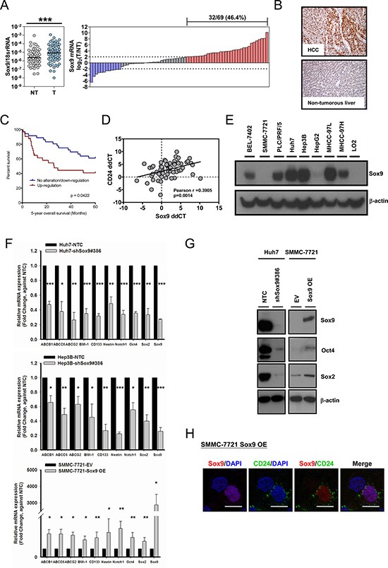
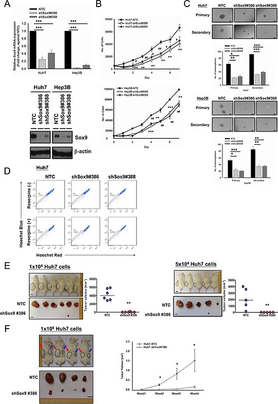
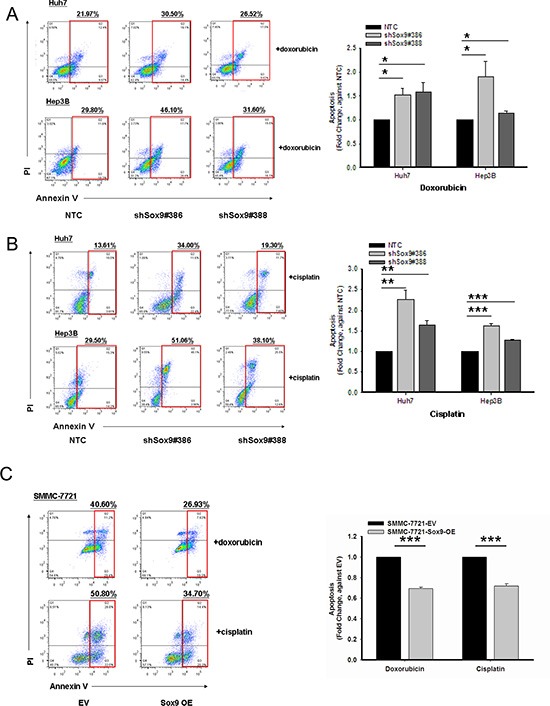
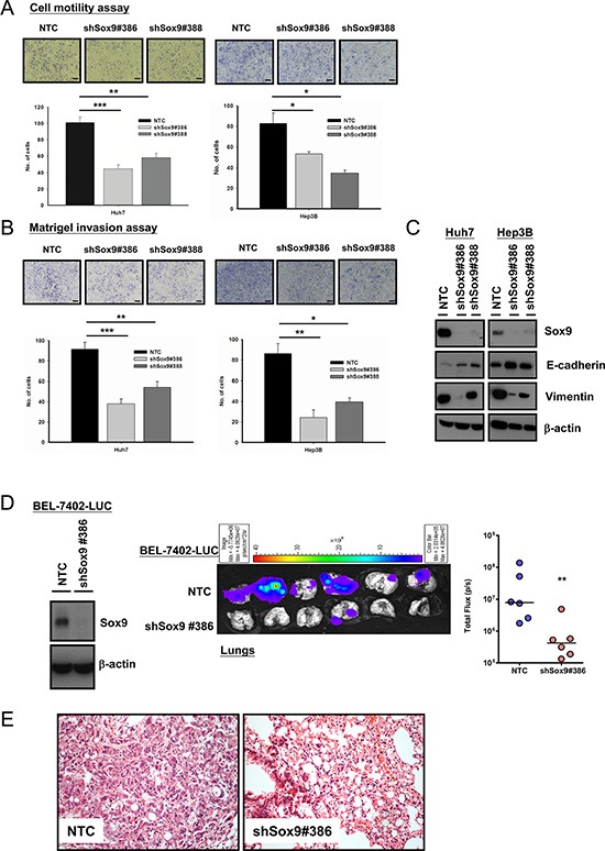
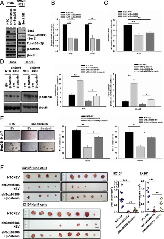
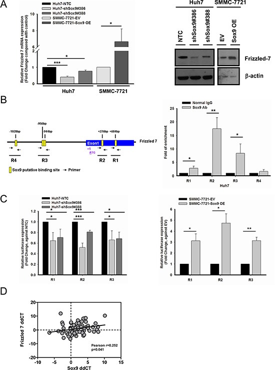
Sox9, an SRY-related HMG box transcription factor, is a progenitor/precursor cell marker of the liver expressed during embryogenesis and following liver injury. In this study, we investigated the role of Sox9 and its molecular mechanism with reference to stemness properties in hepatocellular carcinoma (HCC). Here, we observed upregulation of Sox9 in human HCC tissues compared with the non-tumorous liver counterparts (p < 0.001). Upregulation of Sox9 transcript level was associated with poorer tumor cell differentiation (p = 0.003), venous invasion (p = 0.026), advanced tumor stage (p = 0.044) and shorter overall survival (p = 0.042). Transcript levels of Sox9 and CD24 were positively correlated. Silencing of Sox9 in HCC cells inhibited in vitro cell proliferation and tumorsphere formation, sensitized HCC cells to chemotherapeutic agents, and suppressed in vivo tumorigenicity. In addition, knockdown of Sox9 suppressed HCC cell migration, invasion, and in vivo lung metastasis. Further studies showed that Sox9 endowed stemness features through activation of Wnt/β-catenin signaling, which was confirmed by the partial rescue effect on tumorigenicity and self-renewal upon transfection of active β-catenin in Sox9 knockdown cells. By ChIP and luciferase promoter assays, Frizzled-7 was identified to be the direct transcriptional target of Sox9. In conclusion, Sox9 confers stemness properties of HCC through Frizzled-7 mediated Wnt/β-catenin pathway.

Keywords: Sox9; liver cancer; tumor-initiating cells.


Conflict of interest statement

Nothing to declare.

Figures

Figure 1
Figure 1. Sox9 is upregulated in human HCC and Sox9 expression is associated with expression of stemness markers in vitro
(A) Sox9 was significantly upregulated in HCC tumor (T) versus non-tumorous liver (NT) tissues by qPCR (n = 69; ***p < 0.001, Wilcoxon signed-rank test). Upregulation of Sox9 was found in (32/69) 46.4% of primary HCC cases. Data were presented as the log2 ratio of Sox9 mRNA level in HCC tissues, as compared to the corresponding non-tumorous liver tissues. Upregulation was defined as log2 (T/NT) ≥ 2. (B) Representative images of immunohistochemical staining for Sox9 in clinical HCC samples demonstrated the overexpression at protein level in HCC tissues, while the non-tumorous liver counterparts showed no staining among the hepatocytes. (C) Kaplan-Meier survival curve of overall survival in HCC patients with reference to Sox9 expression. Patients with Sox9 upregulation had lower overall 5-year overall survival rate (p = 0.0422, Kaplan-Meir Log-rank test). (D) Expressions of CD24 and Sox9 were positively correlated in clinical HCC samples. Expression level of CD24 and Sox9 in HCC samples was represented by ddCT (Tumor 18srRNA CT - Gene of interest CT Non-tumor 18srRNA CT Gene of interest CT) (n = 64, Pearson r = 0.3905, p < 0.01, Pearson's correlation). (E) Sox9 was expressed in multiple HCC cell lines by Western blotting, including BEL-7402, PLC/PRF/5, Huh7, Hep3B, MHCC-97L and MHCC-97H. It was not detectable in immortalized liver cell line LO2. (F) Knockdown of Sox9 (clone #386) in Huh7 and Hep3B diminished the expression of multiple chemoresistance-related and stemness-associated genes when compared with non-target control (NTC), while overexpression of (OE) Sox9 in SMMC-7721 reversed the expressions compared with empty vector group (EV) (n = 3, *p < 0.05, **p < 0.01 & ***p < 0.001, t test). The data were presented as mean ± SD. (G) Protein level of two stemness genes, Oct4 and Sox2, were suppressed in Huh7 upon silencing of Sox9 and enhanced upon forced-expression of Sox9 in SMMC-7721. (H) CD24 expression was enhanced upon transient Sox9 overexpression in SMMC-7721 while it was not expressed in untransfected cell (without Sox9 signal) by immunofluorescent staining (Scale bar: 50 μm).
Figure 2
Figure 2. Silencing of Sox9 inhibits cell proliferation, tumorsphere formation and in vivo tumorigenicity in HCC
(A) Two Sox9 stable-knockdown clones (#386 and #388) were established in Huh7 and Hep3B using lentiviral-based shRNA approach. Both Sox9 mRNA (n = 3, ***p < 0.001, t test) and protein levels were suppressed. (B) Knockdown of Sox9 inhibited cell proliferation in both Huh7 and Hep3B cells, when compared with NTC (n = 3, # p < 0.05, **/## p < 0.01 & ***p < 0.001, t test). (C) Knockdown of Sox9 reduced the number and size of the primary and secondary tumorspheres comparing with non-target control (NTC) in both Huh7 and Hep3B cell lines at Day 10 and Day 15, respectively (n = 3, *p < 0.05, **p < 0.01 & ***p < 0.001, t test; Scale bar: 50 μm). The data were presented as mean ± SD. (D) Knockdown of Sox9 suppressed the side population (SP, shSox9#386: 0.137% & shSox9#388: 0.322%) when compared with non-target control (NTC) (1.12%) using Hoechst 33342 labeling. Reserpine sensitivity (lower panel) was used to verify the SPs and to determine optimal staining conditions. (E) Silencing of Sox9 suppressed tumorigenicity in vivo in limited dilution manner of 1 × 10⁴ and 5 × 10⁴ Huh7 cells. Knockdown of Sox9 decreased tumor volume compared with non-target control (NTC) group (Black dotted circle: NTC; **p < 0.01, Mann-Whitney's U test; Scale bar: 1 cm). Suppression of Sox9 showed a decrease in tumor incidence with both cell numbers injected. (F) Huh7 NTC and Huh7 shSox9 cells were injected subcutaneously into NOD/SCID mice (4 mice per group, 1 × 10⁶ cells injected per mouse). Mice were sacrificed at week 4. Knockdown of Sox9 significantly suppressed HCC tumor growth and volume comparing with non-target control (NTC) group (Red arrow: NTC; Blue arrow: shSox9#386; *p < 0.05; Scale bar: 1 cm). The data were presented as mean ± SD.
Figure 3
Figure 3. Sox9 confers chemoresistance in HCC
Cells with stably knockdown of Sox9 were more chemosensitive to both (A) doxorubicin at 0.5 μg/mL for Huh7 and 1.5 μg/mL for Hep3B (n = 3, *p < 0.05, t test), and (B) cisplatin at 5 μg/mL for Huh7 and 10 μg/mL for Hep3B when compared with non-target control (NTC) group after 24-hour treatment at serum free condition (n = 3, **p < 0.01 & ***p < 0.001, t test). Representative Annexin V-PI flow analysis plots of each treatment were shown. (C) Sox9-overexpressing SMMC-7721 cells were more chemoresistant to both doxorubicin at 1 μg/mL and cisplatin 5 μg/mL when compared with empty vector (EV) group (n = 3, ***p < 0.001, t test). Representative Annexin V-PI flow analysis plots of each treatment were shown.
Figure 4
Figure 4. Silencing of Sox9 inhibits cell migration, invasion in vitro and metastasis of HCC in vivo
Knockdown of Sox9 inhibited (A) cell migration ability and (B) invasiveness in both Huh7 and Hep3B cells, when compared with non-target control (NTC) (n = 3, *p < 0.05, **p < 0.01 & ***p < 0.001, t test; Scale bar: 50 μm). (C) Protein expression of E-cadherin was upregulated while vimentin was downregulated upon silencing of Sox9 as demonstrated by Western blotting, signifying inhibition of epithelial-mesenchymal transition with knockdown of Sox9. (D) Sox9 stable-knockdown clone and NTC were established in BEL-7402 luciferase-labeled cells using lentiviral-based shRNA approach. The efficiency of Sox9 silencing was confirmed by Western blotting. Ex vivo xenogen imaging of lungs at 12th week upon injection of either Sox9 silenced or NTC luciferase-labeled BEL-7402 through tail veins of NOD/SCID mice (6 mice per group, 1 × 10⁶ cells injected per mouse). Quantification of luciferase signals showed significant difference between the shSox9 and NTC groups (**p < 0.01, Mann-Whitney's U test). (E) Hematoxylin and eosin staining of mouse lung tissues from NTC and shSox9#386 groups indicated the suppression of pulmonary metastasis upon silencing of Sox9. Representative images were shown (Magnification: 200×).
Figure 5
Figure 5. Sox9 enhances stemness features of HCC through Wnt/β-catenin signaling
(A) Silencing of Sox9 downregulated the expression of phosphorylated (Ser9)-GSK3β and total β-catenin in Huh7 using Western blotting. The reversed effects were observed upon Sox9 overexpression in SMMC-7721. (B) Sox9 silencing (Huh7) suppressed the mRNA levels of c-myc and axin2. (n = 3, *p < 0.05, **p < 0.01 & ***p < 0.001, t test). (C) Knockdown of Sox9 suppressed the activation of Wnt/β-catenin signaling cascade in Huh7 using TOP/FOPFLASH luciferase assay (n = 3, **p < 0.01, t test). (D) Stable expression of β-catenin was enforced in shSox9 clone (#386) in Huh7 and Hep3B and validated using Western blotting. The suppression of Wnt/β-catenin signaling activity was partially rescued in shSox9 knockdown cells upon enforcement of β-catenin when compared with empty vector (EV) group (n = 3, *p < 0.05, **p < 0.01, t test). (E) The suppression of tumorsphere formation ability through knockdown of Sox9 was partially rescued upon β-catenin enforcement (n = 3, *p < 0.05, **p < 0.01, t test; Scale bar: 100 μm). The data were presented as mean ± SD. (F) β-catenin or EV-expressing-shSox9 silencing and EV-expressing Huh7-NTC were subcutaneously inoculated in NOD/SCID mice using 5 × 10³ and 1 × 10⁴ cells for 70 and 60 days, respectively. Upon β-catenin enforcement, the tumorigenicity of Sox9 silenced cells was partially rescued (n = 9, results from two independent experiments for 5 × 10³ group; n = 8 for 1 × 10⁴ group, **p < 0.01 & ***p < 0.001, Mann-Whitney's U test; Scale bar: 1 cm).
Figure 6
Figure 6. Frizzled-7 (FZD7) is a transcriptional target of Sox9 in HCC
(A) FZD7 was suppressed at mRNA (n = 3, *p < 0.05 & ***p < 0.001, t test) and protein levels in Sox9 knockdown Huh7. The reversed effects were observed upon Sox9 overexpression in SMMC-7721. The data were presented as mean ± SD. (B) Chromatin immunoprecipitation (ChIP) assay using Sox9 antibody and normal rabbit IgG in Huh7. Schematic diagram indicated the putative binding sites of Sox9 (+684 bp as R1, +276 bp as R2, −944 bp & −956 bp as R3, −1928 bp as R4) on FZD7 promoter and exon 1 region (Yellow box: Sox9 putative binding site; Black arrow: primer for ChIP assay). Sox9 bound to R1, R2 and R3, but not R4 in Huh7 cells (n = 3, *p < 0.05 & **p < 0.01, t test). (C) A significant decrease of signals was detected in silenced Sox9 Huh7 comparing with non-target control (NTC) group, while the signals were upregulated in Sox9-overexpressing SMMC-7721 comparing with EV transfected group using luciferase reporter assay in R1, R2 and R3. (n = 3, *p < 0.05, **p < 0.01 & ***p < 0.001, t test). The data were presented as mean ± SD. (D) A positive correlation between FZD7 and Sox9 mRNA expressions was found in clinical HCC samples. Expression level of FZD7 and Sox9 in paired HCC samples was represented by ddCT (Tumor 18srRNA CT - Gene of interest CT- Non-tumor 18srRNA CT - Gene of interest CT) (n = 66, Pearson r = 0.252, p < 0.05, Pearson's correlation).

Similar articles

Cited by

References

    1. Lefebvre V, Dumitriu B, Penzo-Mendez A, Han Y, Pallavi B. Control of cell fate and differentiation by Sry-related high-mobility-group box (Sox) transcription factors. Int J Biochem Cell Biol. 2007;39:2195–214. - PMC - PubMed
    1. Furuyama K, Kawaguchi Y, Akiyama H, Horiguchi M, Kodama S, Kuhara T, Hosokawa S, Elbahrawy A, Soeda T, Koizumi M, Masui T, Kawaguchi M, Takaori K, et al. Continuous cell supply from a Sox9-expressing progenitor zone in adult liver, exocrine pancreas and intestine. Nat Genet. 2011;43:34–41. - PubMed
    1. Yimlamai D, Christodoulou C, Galli GG, Yanger K, Pepe-Mooney B, Gurung B, Shrestha K, Cahan P, Stanger BZ, Camargo FD. Hippo pathway activity influences liver cell fate. Cell. 2014;157:1324–38. - PMC - PubMed
    1. Zhong WD, Qin GQ, Dai QS, Han ZD, Chen SM, Ling XH, Fu X, Cai C, Chen JH, Chen XB, Lin ZY, Deng YH, Wu SL, et al. SOXs in human prostate cancer: implication as progression and prognosis factors. BMC Cancer. 2012;12:248. - PMC - PubMed
    1. Zhou CH, Ye LP, Ye SX, Li Y, Zhang XY, Xu XY, Gong LY. Clinical significance of SOX9 in human non-small cell lung cancer progression and overall patient survival. J Exp Clin Cancer Res. 2012;31:18. - PMC - PubMed

MeSH terms