

Stabilin-1 is expressed in human breast cancer and supports tumor growth in mammary adenocarcinoma mouse model
- PMID: 27105498
- PMCID: PMC5058742
- DOI: 10.18632/oncotarget.8857
Stabilin-1 is expressed in human breast cancer and supports tumor growth in mammary adenocarcinoma mouse model
Abstract
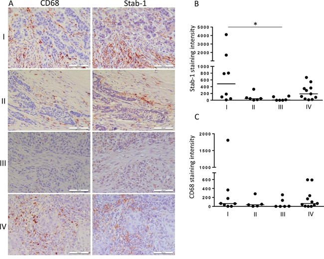
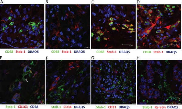
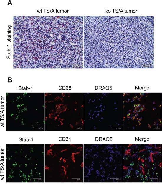
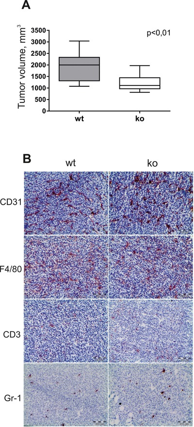
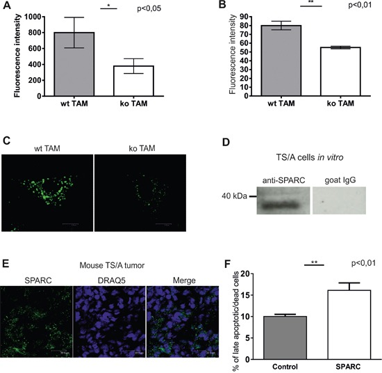
Stabilin-1 is a multifunctional scavenger receptor expressed on alternatively-activated macrophages. Stabilin-1 mediates phagocytosis of "unwanted-self" components, intracellular sorting, and endocytic clearance of extracellular ligands including SPARC that modulates breast cancer growth. The expression of stabilin-1 was found on tumor-associated macrophages (TAM) in mouse and human cancers including melanoma, lymphoma, glioblastoma, and pancreatic insulinoma. Despite its tumor-promoting role in mouse models of melanoma and lymphoma the expression and functional role of stabilin-1 in breast cancer was unknown. Here, we demonstrate that stabilin-1 is expressed on TAM in human breast cancer, and its expression is most pronounced on stage I disease. Using stabilin-1 knockout (ko) mice we show that stabilin-1 facilitates growth of mouse TS/A mammary adenocarcinoma. Endocytosis assay on stabilin-1 ko TAM demonstrated impaired clearance of stabilin-1 ligands including SPARC that was capable of inducing cell death in TS/A cells. Affymetrix microarray analysis on purified TAM and reporter assays in stabilin-1 expressing cell lines demonstrated no influence of stabilin-1 expression on intracellular signalling. Our results suggest stabilin-1 mediated silent clearance of extracellular tumor growth-inhibiting factors (e.g. SPARC) as a mechanism of stabilin-1 induced tumor growth. Silent clearance function of stabilin-1 makes it an attractive candidate for delivery of immunomodulatory anti-cancer therapeutic drugs to TAM.
Keywords: SPARC; breast cancer; scavenger receptor; stabilin-1; tumor-associated macrophages.
Conflict of interest statement
The authors declare that there are no conflicts of interest.
Figures

Similar articles
-
CD68+, but not stabilin-1+ tumor associated macrophages in gaps of ductal tumor structures negatively correlate with the lymphatic metastasis in human breast cancer.Immunobiology. 2017 Jan;222(1):31-38. doi: 10.1016/j.imbio.2015.09.011. Epub 2015 Sep 8. Immunobiology. 2017. PMID: 26391151
-
Novel function of alternatively activated macrophages: stabilin-1-mediated clearance of SPARC.J Immunol. 2006 May 15;176(10):5825-32. doi: 10.4049/jimmunol.176.10.5825. J Immunol. 2006. PMID: 16670288
-
Stabilin-1, a homeostatic scavenger receptor with multiple functions.J Cell Mol Med. 2006 Jul-Sep;10(3):635-49. doi: 10.1111/j.1582-4934.2006.tb00425.x. J Cell Mol Med. 2006. PMID: 16989725 Free PMC article. Review.
-
Identification of a sequence in the matricellular protein SPARC that interacts with the scavenger receptor stabilin-1.J Cell Biochem. 2011 Apr;112(4):1003-8. doi: 10.1002/jcb.23015. J Cell Biochem. 2011. PMID: 21308731
-
Multifunctional receptor stabilin-1 in homeostasis and disease.ScientificWorldJournal. 2010 Oct 12;10:2039-53. doi: 10.1100/tsw.2010.189. ScientificWorldJournal. 2010. PMID: 20953554 Free PMC article. Review.
Cited by
-
The relationship between stromal cell derived SPARC in human gastric cancer tissue and its clinicopathologic significance.Oncotarget. 2017 Sep 21;8(49):86240-86252. doi: 10.18632/oncotarget.21133. eCollection 2017 Oct 17. Oncotarget. 2017. PMID: 29156791 Free PMC article.
-
Scavenger Receptors: Novel Roles in the Pathogenesis of Liver Inflammation and Cancer.Semin Liver Dis. 2022 Feb;42(1):61-76. doi: 10.1055/s-0041-1733876. Epub 2021 Sep 22. Semin Liver Dis. 2022. PMID: 34553345 Free PMC article. Review.
-
Targeting of TAMs: can we be more clever than cancer cells?Cell Mol Immunol. 2024 Dec;21(12):1376-1409. doi: 10.1038/s41423-024-01232-z. Epub 2024 Nov 8. Cell Mol Immunol. 2024. PMID: 39516356 Free PMC article. Review.
-
The Role of Stabilin-1 in Lymphocyte Trafficking and Macrophage Scavenging in the Liver Microenvironment.Biomolecules. 2019 Jul 16;9(7):283. doi: 10.3390/biom9070283. Biomolecules. 2019. PMID: 31315308 Free PMC article. Review.
-
STAB1 Promotes Acute Myeloid Leukemia Progression by Activating the IKK/NF-κB Pathway and Increasing M2 Macrophage Polarization.Cancer Sci. 2025 Jun;116(6):1508-1521. doi: 10.1111/cas.70044. Epub 2025 Mar 13. Cancer Sci. 2025. PMID: 40083109 Free PMC article.
References
-
- Schledzewski K, Geraud C, Arnold B, Wang S, Grone HJ, Kempf T, Wollert KC, Straub BK, Schirmacher P, Demory A, Schonhaber H, Gratchev A, Dietz L, Thierse HJ, Kzhyshkowska J, Goerdt S. Deficiency of liver sinusoidal scavenger receptors stabilin-1 and -2 in mice causes glomerulofibrotic nephropathy via impaired hepatic clearance of noxious blood factors. The Journal of clinical investigation. 2011;121:703–714. - PMC - PubMed
-
- Park SY, Jung MY, Lee SJ, Kang KB, Gratchev A, Riabov V, Kzhyshkowska J, Kim IS. Stabilin-1 mediates phosphatidylserine-dependent clearance of cell corpses in alternatively activated macrophages. Journal of cell science. 2009;122:3365–3373. - PubMed
-
- Kzhyshkowska J, Mamidi S, Gratchev A, Kremmer E, Schmuttermaier C, Krusell L, Haus G, Utikal J, Schledzewski K, Scholtze J, Goerdt S. Novel stabilin-1 interacting chitinase-like protein (SI-CLP) is up-regulated in alternatively activated macrophages and secreted via lysosomal pathway. Blood. 2006;107:3221–3228. - PubMed
MeSH terms
Substances
LinkOut - more resources
Full Text Sources
Other Literature Sources
Medical
Molecular Biology Databases
Research Materials
Miscellaneous
