. 2016 May 17;7(20):29677-88.
doi: 10.18632/oncotarget.8820.

Gene expression profiling of normal thyroid tissue from patients with thyroid carcinoma

Affiliations

Gene expression profiling of normal thyroid tissue from patients with thyroid carcinoma

Roberto Ria et al. Oncotarget. .

Abstract

Gene expression profiling (GEP) of normal thyroid tissue from 43 patients with thyroid carcinoma, 6 with thyroid adenoma, 42 with multinodular goiter, and 6 with Graves-Basedow disease was carried out with the aim of achieving a better understanding of the genetic mechanisms underlying the role of normal cells surrounding the tumor in the thyroid cancer progression. Unsupervised and supervised analyses were performed to compare samples from neoplastic and non-neoplastic diseases. GEP and subsequent RT-PCR analysis identified 28 differentially expressed genes. Functional assessment revealed that they are involved in tumorigenesis and cancer progression. The distinct GEP is likely to reflect the onset and/or progression of thyroid cancer, its molecular classification, and the identification of new potential prognostic factors, thus allowing to pinpoint selective gene targets with the aim of realizing more precise preoperative diagnostic procedures and novel therapeutic approaches.This study is focused on the gene expression profiling analysis followed by RT-PCR of normal thyroid tissues from patients with neoplastic and non-neoplastic thyroid diseases. Twenty-eight genes were found to be differentially expressed in normal cells surrounding the tumor in the thyroid cancer. The genes dysregulated in normal tissue samples from patients with thyroid tumors may represent new molecular markers, useful for their diagnostic, prognostic and possibly therapeutic implications.

Keywords: gene expression profile; hypoxia; microenvironment; oncogenes; thyroid cancer.


Conflict of interest statement

All the authors declare no conflict of interests.

Figures

Figure 1
Figure 1. Evaluation of neoplastic infiltration
Ematoxylin/eosin staining showing non neoplastic thyroid tissues in panel A. versus neoplastic infiltration in panel B. Immunohistochemical staining for Cytokeratin 19: in panel C. CK19 is not detected in normal tissue, while diffuse immunoreactivity for CK19 in a papillary thyroid cancer infiltration is visible in panel D.
Figure 2
Figure 2. Unsupervised analysis of gene expression profiles in normal tissue of tumoral and non-tumoral thyroids
The dendrogram was generated with a hierarchical clustering algorithm based on the average-linkage method. In the matrix, each column represents a sample, and each row a gene.
Figure 3
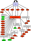
Figure 3. Global role of up- and down-regulated genes in thyroid cancer development and progression
Correlation of up- (red-labeled) and down-regulated (green-labeled) studied genes and their corresponding biological functions with tumor progression.
Figure 4
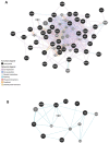
Figure 4. Gene network analysis in normal tissue of tumoral thyroids
A. General analysis was based on protein-protein interaction and pathway databases with the nucleocytoplasmic transport-related genes (1.29-fold; P,0.05). GeneMANIA retrieved known and predicted interactions between these genes and added extra genes (grey circles) that are strongly connected to query genes (dark circles). B. Functional gene network in normal tissue of patients with thyroid cancer. Correlation of the three genes, namely HIF1A, TUFT1, BHLHB2, involved in angiogenesis and response to hypoxya with other disregulated genes, particularly JUND.
Figure 5
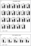
Figure 5. Measurement of gene expression by real-time RT-PCR
A. Significant increase in normal tissues from neoplastic vs non-neoplastic thyroids of ZFP36L1, TUFT1, SLCA2A3, SKP1, RIPK5, RAB7B, PTGS2, KLF6, JUND, IRF1, IER3, HIF1A, HBEGF, GBP1, GADD45A, GADD45B, EIF4A3, DUSP5, CCNL1, CADM1, BMP2, BHLHB2, AUXD1. B. In contrast, only 5 genes were down-regulated in these tissues: WDR48, TEF, PNPLA7, ACCS, and KLK-4. Values are expressed as mean ± 1SD for 32 normal tissues from neoplastic thyroids and 28 non-neoplastic thyroids. Significance of changes was calculated by the Wilcoxon-Wilcox test.
Figure 6
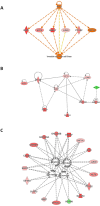
Figure 6. Gene interaction network based on information from the Ingenuity Pathways Knowledge Base under the IFNG control
A. Correlation with cancer cell invasion. B. Gene interaction network based on information from the Ingenuity Pathways Knowledge Base under the HIF1A control. Genes that are up- or down-regulated are labelled in red and green, respectively. C. Gene interaction network based on information from the Ingenuity Pathways Knowledge Base related to cell survival. Correlation of genes dysregulated in tumoral thyroids with cell viability, survival, death and apoptosis. Genes that are up- or down-regulated are labelled in red and green, respectively.

References

    1. Shirazi HA, Hedayati M, Daneshpour MS, Shafiee A, Azizi F. Analysis of loss of heterozygosity effect on thyroid tumor with oxyphilia cell locus in familial non medullar y thyroid carcinoma in Iranian families. Indian J Hum Genet. 2012;18:340–3. - PMC - PubMed
    1. Ghossein R. Problems and controversies in the histopathology of thyroid carcinomas of follicular cell origin. Arch Pathol Lab Med. 2009;133:683–91. - PubMed
    1. Fassnacht M, Kreissl MC, Weismann D, Allolio B. New targets and therapeutic approaches for endocrine malignancies. Pharmacol Ther. 2009;123:117–41. - PubMed
    1. Hedayati M, Zarif Yeganeh M, Sheik hol Eslami S, Rezghi Barez S, Hoghooghi Rad L, Azizi F. Predominant RET germline mutations in exons 10, 11, and 16 in Iranian patients with hereditary medullary thyroid carcinoma. J Thyroid Res. 2011:264248–2011. - PMC - PubMed
    1. Kondo T, Ezzat S, Asa SL. Pathogenetic mechanisms in thyroid follicular-cell neoplasia. Nat Rev Cancer. 2006;6:292–306. - PubMed

Substances