. 2016 Sep 6;44(15):7189-203.
doi: 10.1093/nar/gkw331. Epub 2016 Apr 22.

Establishment of a promoter-based chromatin architecture on recently replicated DNA can accommodate variable inter-nucleosome spacing

Affiliations

Establishment of a promoter-based chromatin architecture on recently replicated DNA can accommodate variable inter-nucleosome spacing

Ross T Fennessy et al. Nucleic Acids Res. .

Abstract

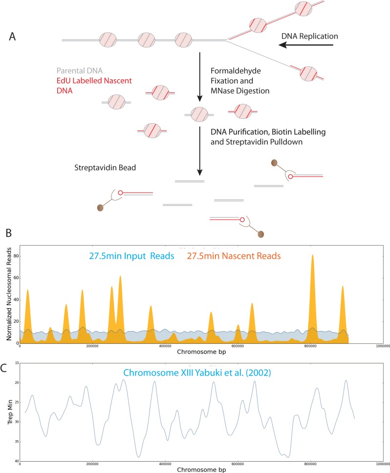
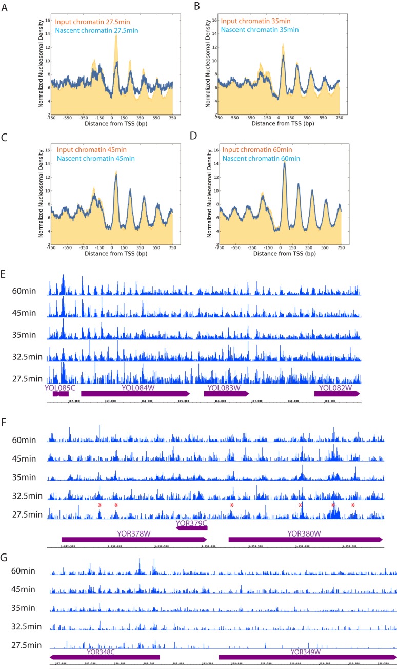
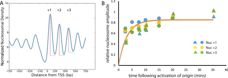
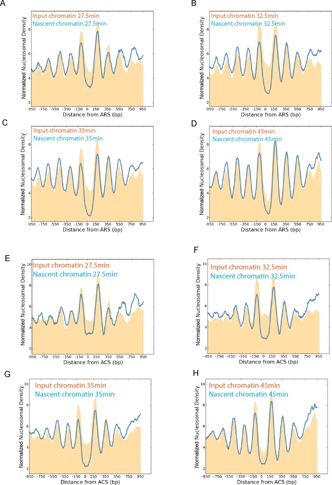
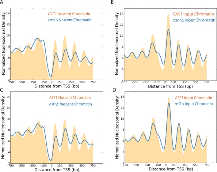
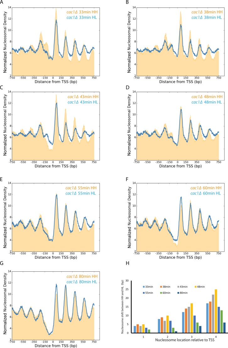
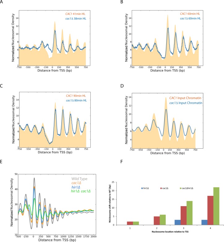
Nucleosomes, the fundamental subunits of eukaryotic chromatin, are organized with respect to transcriptional start sites. A major challenge to the persistence of this organization is the disassembly of nucleosomes during DNA replication. Here, we use complimentary approaches to map the locations of nucleosomes on recently replicated DNA. We find that nucleosomes are substantially realigned with promoters during the minutes following DNA replication. As a result, the nucleosomal landscape is largely re-established before newly replicated chromosomes are partitioned into daughter cells and can serve as a platform for the re-establishment of gene expression programmes. When the supply of histones is disrupted through mutation of the chaperone Caf1, a promoter-based architecture is generated, but with increased inter-nucleosomal spacing. This indicates that the chromatin remodelling enzymes responsible for spacing nucleosomes are capable of organizing nucleosomes with a range of different linker DNA lengths.


Figures

Figure 1.
Figure 1.
A system for isolation of nascent chromatin by EdU labelling of newly replicated DNA. (A) Schematic illustration of the EdU approach for isolation of nascent nucleosomal DNA. (B) Reads for replicating (nascent: orange) and unreplicated (input: blue) nucleosomal DNA per bp along chromosome 13 for an early S-phase time point, 27.5 min post-release from G1 arrest. (C) Replication profiles from previously annotated origins of replication for chromosome 13 identified by S-phase copy number (43).
Figure 2.
Figure 2.
Characterization of nascent chromatin by EdU labelling of newly replicated DNA. Normalized frequency of nucleosome dyads aligned to the TSS of all genes (n = 5015) at the indicated time points following release from G1 arrest. The distribution of replicated fragments (nascent: blue) isolated by affinity purification of EdU labelled fragments is shown in comparison to the total chromatin isolated prior to pull down (input: orange) (A) 27.5, (B) 35, (C) 45, (D) 60 min following release from α factor arrest. Reads from EdU enriched chromatin isolated at the time points indicated following G1 arrest are shown across individual loci in (E and F). Across the locus shown in (E) many chromatin features are distinguishable at 27.5 min and the greatest maturation occurs between 27.5 and 32.5 min consistent with what is observed in the average profile of all genes. (F) Shows a locus at which many nucleosomes are less well defined and some chromatin features (indicated with a red asterisk) are detectable at the earliest time point and do not change or disperse over the time course. (G) This region is replicated later and as a result is depleted for reads isolated from early S-phase chromatin.
Figure 3.
Figure 3.
Kinetics of nucleosome organization. The depth of the oscillation in nucleosomal read depth was determined for the +1, +2 and +3 nucleosomes as indicated in (A). The oscillation depth in nascent chromatin at nuc +1 (blue), nuc +2 (yellow), nuc +3 (green) was then expressed as a fraction of that observed in the input chromatin for two repeats of an EdU time course at the time points indicated (B). The time values were calculated based on the distribution of replicated fragments observed at origins multiplied by the rate of elongation for DNA polymerase, 1.6 kb/min (17). Time points for two biological repeats are shown as circles and triangles. A fit to the first order rate equation, y = Ae−(kt) is shown (orange) which allows estimation of the half time for nucleosome positioning as 2.1 min. The residual, R2, for this fit is 0.48.
Figure 4.
Figure 4.
Chromatin maturation at origins of DNA replication. Chromatin from the EdU enrichment time course was plotted with respect to 205 replication origins (46). Nascent (blue) and input chromatin (orange) are plotted 27.5 (A), 32.5 (B), 35 (C) and 45 min (D) following release from G1 arrest. The +2 and +3 Nucleosomes are significantly ordered at the first time point and this improves over the following minutes. As many replication origins are located adjacent to transcribed genes, the same analysis was performed with 127 replication origins for which no TSS was present within 500 bp of the origin (E–H). Nucleosomes are not as precisely aligned to TSS-free origins in comparison to all origins (Compare input chromatin A–D to that of E–H). In particular the nucleosome depleted region at origins is poorly defined in early S-phase. Organization of the +2 and +3 nucleosomes at replication origins with no adjacent TSS mature at a similar rate to that observed at all origins.
Figure 5.
Figure 5.
Loss of histone chaperones perturbs nascent chromatin organization. Normalized frequency of nascent nucleosomal dyads aligned to the TSS of all genes (n = 5015) in wild-type (orange), cac1Δ (blue) (A) and asf1Δ (blue) (C) deficient strains, 10 min following addition of EdU to an asynchronously growing culture. The normalized frequency of input chromatin prior to enrichment for newly replicated fragments for wild-type, cac1Δ (B) and asf1Δ strains (D).
Figure 6.
Figure 6.
Alteration of nucleosome spacing in nascent chromatin in the absence of Cac1. Isolation of nascent DNA by isotope labelling and caesium chloride gradient density separation provides a means to track the relatively slow maturation of chromatin in a cac1Δ mutant. Normalized frequency of nucleosomal dyads aligned to the TSS for replicated HL (blue) and unreplciated HH (orange) labelled nucleosomal fragments isolated 33 (A), 38 (B), 43 (C), 48 (D), 55 (E), 60 (F) and 80 min (G) following release from G1 arrest. Quantitation of the 3′ shift in the average nucleosome location for +1, +2, +3 and +4 nucleosomes for each time point is shown in (H).
Figure 7.
Figure 7.
The defect to nucleosome spacing in the absence of Cac1 is restored post-replication and enhanced in the absence of replication-independent histone turnover. Alignment of nucleosomal reads on nascent DNA to the TSS in wild-type (orange) and cac1Δ strains (blue) illustrates progressive accumulation of a spacing defect in mid S-phase (A and B). This is restored in late S-phase (C). The spacing defect in asynchronous total chromatin (D) is less than that observed in mid S-phase (A and B). Nucleosomal reads from asynchronous wild-type, cac1Δ, hir1Δ and hir1Δcac1Δ strains were aligned to the TSS of all genes (n = 5015) (E). The nucleosome depleted region at promoters is partially filled in a hir1Δcac1Δ strain (green) in comparison to cac1Δ (orange), hir1Δ (blue) and wild-type (grey). The defect to nucleosome spacing is quantified in (F). The defect is increased in the hir1Δcac1Δ consistent with replication-independent histone turnover acting to restore nucleosome density on coding regions.

References

    1. Liu C.L., Kaplan T., Kim M., Buratowski S., Schreiber S.L., Friedman N., Rando O.J. Single-nucleosome mapping of histone modifications in S. cerevisiae. PLoS Biol. 2005;3:e328. - PMC - PubMed
    1. Filion G.J., van Bemmel J.G., Braunschweig U., Talhout W., Kind J., Ward L.D., Brugman W., de Castro I.J., Kerkhoven R.M., Bussemaker H.J., et al. Systematic protein location mapping reveals five principal chromatin types in Drosophila cells. Cell. 2010;143:212–224. - PMC - PubMed
    1. Hoffman M.M., Ernst J., Wilder S.P., Kundaje A., Harris R.S., Libbrecht M., Giardine B., Ellenbogen P.M., Bilmes J.A., Birney E., et al. Integrative annotation of chromatin elements from ENCODE data. Nucleic Acids Res. 2013;41:827–841. - PMC - PubMed
    1. Alabert C., Barth T.K., Reveron-Gomez N., Sidoli S., Schmidt A., Jensen O.N., Imhof A., Groth A. Two distinct modes for propagation of histone PTMs across the cell cycle. Genes Dev. 2015;29:585–590. - PMC - PubMed
    1. Faure A.J., Schmidt D., Watt S., Schwalie P.C., Wilson M.D., Xu H., Ramsay R.G., Odom D.T., Flicek P. Cohesin regulates tissue-specific expression by stabilizing highly occupied cis-regulatory modules. Genome Res. 2012;22:2163–2175. - PMC - PubMed