. 2016 Jun 1;310(11):L1206-17.
doi: 10.1152/ajplung.00424.2015. Epub 2016 Apr 22.

The vitronectin RGD motif regulates TGF-β-induced alveolar epithelial cell apoptosis

Affiliations

The vitronectin RGD motif regulates TGF-β-induced alveolar epithelial cell apoptosis

Amanda K Wheaton et al. Am J Physiol Lung Cell Mol Physiol. .

Abstract

Transforming growth factor-β (TGF-β) is a critical driver of acute lung injury and fibrosis. Injury leads to activation of TGF-β, which regulates changes in the cellular and matrix makeup of the lung during the repair and fibrosis phase. TGF-β can also initiate alveolar epithelial cell (AEC) apoptosis. Injury leads to destruction of the laminin-rich basement membrane, which is replaced by a provisional matrix composed of arginine-glycine-aspartate (RGD) motif-containing plasma matrix proteins, including vitronectin and fibronectin. To determine the role of specific matrix proteins on TGF-β-induced apoptosis, we studied primary AECs cultured on different matrix conditions and utilized mice with deletion of vitronectin (Vtn(-/-)) or mice in which the vitronectin RGD motif is mutated to nonintegrin-binding arginine-glycine-glutamate (RGE) (Vtn(RGE/RGE)). We found that AECs cultured on fibronectin and vitronectin or in wild-type mouse serum are resistant to TGF-β-induced apoptosis. In contrast, AECs cultured on laminin or in serum from Vtn(-/-) or Vtn(RGE/RGE) mice undergo robust TGF-β-induced apoptosis. Plasminogen activator inhibitor-1 (PAI-1) sensitizes AECs to greater apoptosis by disrupting AEC engagement to vitronectin. Inhibition of integrin-associated signaling proteins augments AEC apoptosis. Mice with transgenic deletion of PAI-1 have less apoptosis after bleomycin, but deletion of vitronectin or disruption of the vitronectin RGD motif reverses this protection, suggesting that the proapoptotic function of PAI-1 is mediated through vitronectin inhibition. Collectively, these data suggest that integrin-matrix signaling is an important regulator of TGF-β-mediated AEC apoptosis and that PAI-1 functions as a natural regulator of this interaction.

Keywords: apoptosis; epithelial; fibrosis; lung; matrix.


Figures

Fig. 1.
Fig. 1.
Purity of isolated murine alveolar epithelial cells (AECs). Isolated murine AECs were stained with isotype control antibody (A) or antibody to pro-surfactant protein-C (pro-SPC) (B) and fluorescent-conjugated anti-rabbit secondary antibody. Data also shown as histogram overlay (C). SSC, side scatter.
Fig. 2.
Fig. 2.
Extracellular matrix regulates AEC apoptotic response to TGF-β. A–C: TUNEL staining (red) of primary AECs cultured on Matrigel (Mg) (A), fibronectin (Fn) (B) or vitronectin (Vtn) (C) and treated with transforming growth factor-β (TGF-β) (4 ng/ml) for 48 h. D: quantification of TUNEL-positive cells; n = 5, *P < 0.05 compared with AECs on Mg. E: activation of caspase-3/7 by AECs cultured on Mg, Fn, or Vtn treated with or without TGF-β (4 ng/ml) for 24 h. Values are expressed as fold differences in relative luminescent units; n = 5, *P < 0.05 compared with AECs on Mg.
Fig. 3.
Fig. 3.
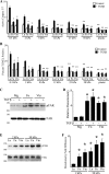
Composition and rigidity of the matrix regulate AEC apoptosis in response to TGF-β. A and B: AECs were cultured on hydrogels of defined stiffness (Matrigen Life Technologies) coated with laminin (Ln), Fn, or Vtn (10 μg/ml), and activation of caspase-3/7 (A) and percentage of TUNEL-positive cells (B) in response to TGF-β (4 ng/ml) was determined. The influence of matrix protein composition (*P < 0.05 compared with TGF-β-treated AECs on Ln of equal stiffness) and stiffness (**P < 0.05 compared with TGF-β-treated AECs on similar matrix protein at 0.5-kPa stiffness) was determined, n = 5. C and D: immunoblot for focal adhesion kinase (FAK) and phospho-FAK (pFAK) (C) were quantified by densitometry (D); n = 5, *P < 0.05 compared with AECs on Mg. E and F: immunoblot of FAK and pFAK of AECs cultured on Ln, Fn, or Vtn coated with 2-kPa or 50-kPa stiff matrix (E) and quantified by densitometry (F); n = 5, *P < 0.05 compared with AEC on 2-kPa Ln.
Fig. 4.
Fig. 4.
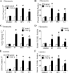
FAK/Src/Rho activation inhibits AEC apoptosis in vitro. Primary AECs cultured on tissue culture plates coated with 10 μg/ml of fibronectin (A and B), vitronectin (C and D), or laminin (E and F) and treated with inhibitors to FAK (10 μM PF573228), Src (10 μM PP2), or Rho kinase (10 μM Y27632) were assayed for activation of caspase-3/7 (A, C, and E) and percentage of TUNEL-positive cells (B, D, and F) in response to TGF-β (4 ng/ml) compared with AECs treated with DMSO vehicle control; n = 6, *P < 0.05 compared with AECs treated with DMSO and TGF-β.
Fig. 5.
Fig. 5.
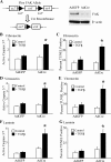
Deletion of FAK augments TGF-β-induced AEC apoptosis. A: immunoblot for FAK from primary AECs from floxed FAK mice after treatment with adenovirus-expressing Cre recombinase (AdCre, 50 pfu/cell) compared with cells treated with control adenovirus-expressing green fluorescent protein (GFP) (AdGFP). Primary floxed FAK AECs treated with AdGFP or AdCre were cultured on tissue culture plates coated with 10 μg/ml fibronectin (B and C), vitronectin (D and E), or laminin (F and G), and activation of caspase-3/7 (B, D, and F) and percentage of TUNEL-positive cells (C, E, and G) in response to TGF-β was determined. *P < 0.05 compared with AECs treated with AdGFP and TGF-β, n = 6.
Fig. 6.
Fig. 6.
Generation of vitronectin arginine-glycine-glutamate (RGE) mice. A: generation of VTNRGE mice in which the integrin-binding arginine-glycine-aspartate (RGD) motif of vitronectin was mutated to an RGE. The targeting vector includes a 3.8-kb 5′ homology arm upstream of the RGD site and 6-kb 3′ homology arm downstream and added FRT flanked neomycin resistance. Resulting mice were crossed with a constitutive FlpE mouse to remove the FRT-neo cassette. B: normal levels of vitronectin in VTNRGE/RGE mice were verified by immunoblot of serum isolated from VTN-null (VTN−/−), wild-type (WT), and VTNRGE/RGE mice.
Fig. 7.
Fig. 7.
Vitronectin RGD motif regulates TGF-β-induced AEC apoptosis. A and B: AECs cultured on tissue culture plates coated with serum from VTN−/− and VTNRGE/RGE mice were assessed for FAK phosphorylation (pFAK) by immunoblot (A) and quantified by densitometry (B); n = 5, *P < 0.05 compared with WT. C and D: AECs cultured on tissue culture plates coated with serum from VTN−/− and VTNRGE/RGE mice were assessed for TGF-β-induced activation of caspase-3/7 (C) and percentage of TUNEL-positive cells (D) compared with AECs cultured on WT serum. *P < 0.05 compared with TGF-β-treated AECs on WT serum, n = 6.
Fig. 8.
Fig. 8.
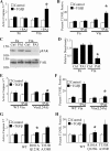
Plasminogen activator inhibitor-1 (PAI) is a natural inhibitor of vitronectin-mediated protection from TGF-β-mediated apoptosis. A and B: AECs cultured on vitronectin (Vtn) and fibronectin (Fn) were treated with PAI-1 (50 ng/ml) and TGF-β, and activation of caspase-3/7 (A) and percentage of TUNEL-positive cells (B) were determined. *P < 0.05 compared with AECs on Fn treated with TGF-β, n = 5. C and D: immunoblot for FAK and phosphorylation of FAK (pFAK) in AECs cultured on Vtn and Fn and treated with PAI-1 and TGF-β (C) and quantified by densitometry (D); n = 5, *P < 0.05 compared with AECs not treated with PAI-1. E and F: AECs cultured on WT Vtn and Vtn(L24A) were treated with PAI-1 with TGF-β-activated caspase-3/7 (E), and the percentage of TUNEL-positive cells (F) was determined; n = 6, *P < 0.05 compared with AECs on WT VTN treated with TGF-β. G and H: activation of caspase-3/7 (G) and percentage of TUNEL-positive cells (H) by AECs cultured on VTN stimulated with TGF-β and WT PAI-1, mutant PAI-1, which does not bind plasminogen activator [PAI-1(T333R,A338R)], and PAI-1 mutant, which does not engage VTN [PAI-1(R101A,Q123K)]; n = 6, *P < 0.05 compared with AECs treated with WT PAI-1 and TGF-β.
Fig. 9.
Fig. 9.
Induction of PAI-1 by bleomycin and TGF-β. A and B: immunoblot (A) for PAI-1 in equal volume of bronchoalveolar lavage (BAL) fluid from uninjured mice or 5 days after bleomycin (2 U/kg) injury demonstrates robust upregulation of PAI-1 after bleomycin, as quantified by densitometry (B); n = 5, P < 0.05 compared with uninjured BAL. BAL from PAI-1-null mice (PAI−/−) is used as a negative control. C and D: AECs cultured on Vtn, Fn, or Mg have increased PAI-1 levels 24 h after TGF-β (4 ng/ml) determined by immunoblot (C) and quantified by densitometry (D); n = 5, *P < 0.05 compared with no TGF-β.
Fig. 10.
Fig. 10.
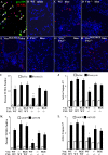
PAI-1 augments AEC apoptosis through inhibition of vitronectin RGD domain in vivo. A: 5-day bleomycin WT mouse lung section was stained for pro-SPC (green) and TUNEL (red). Numerous pro-SPC/TUNEL-copositive cells are demonstrated (arrows), ×600. B–H: 5 days after saline (B) or bleomycin injury, WT (C), vitronectin-null (Vtn−/−) (D), vitronectin RGE (VtnRGE/RGE) (E), PAI-1-null (PAI−/−) (F), vitronectin-null/PAI-1-null (VTN−/−PAI−/−) (G), and vitronectin RGE/PAI-1-null (VTNRGE/RGEPAI−/−) (H) mouse lungs were stained for TUNEL (red), ×200. I: quantification of TUNEL-positive nuclei after bleomycin injury. J: activation of caspase-3/7 from whole lung lysate 5 days after bleomycin. K: quantification of TUNEL-positive nuclei 5 days after treatment with adenovirus-encoding TGF-β (AdTGF-β) or GFP. L: activation of caspase-3/7 from whole-lung lysate 5 days after AdTGF-β or AdGFP. *P < 0.05, n = 5–12 compared with WT mice treated with bleomycin or AdTGF-β.

References

    1. Akiyama SK. Purification of vitronectin. Curr Protoc Cell Biol 60: 16, 2013. - PubMed
    1. Ashley SL, Sisson TH, Wheaton AK, Kim KK, Wilke CA, Ajayi IO, Subbotina N, Wang S, Duckett CS, Moore BB, Horowitz JC. Targeting inhibitor of apoptosis proteins protects from bleomycin-induced lung fibrosis. Am J Respir Cell Mol Biol 54: 482–492, 2016. - PMC - PubMed
    1. Bale MD, Wohlfahrt LA, Mosher DF, Tomasini B, Sutton RC. Identification of vitronectin as a major plasma protein adsorbed on polymer surfaces of different copolymer composition. Blood 74: 2698–2706, 1989. - PubMed
    1. Barnes DW, Silnutzer J. Isolation of human serum spreading factor. J Biol Chem 258: 12548–12552, 1983. - PubMed
    1. Beausejour M, Noel D, Thibodeau S, Bouchard V, Harnois C, Beaulieu JF, Demers MJ, Vachon PH. Integrin/Fak/Src-mediated regulation of cell survival and anoikis in human intestinal epithelial crypt cells: selective engagement and roles of PI3-K isoform complexes. Apoptosis 17: 566–578, 2012. - PMC - PubMed

Publication types