Review
. 2017 Jan 15;595(2):557-570.
doi: 10.1113/JP271021. Epub 2016 May 29.

Enteric glia: the most alimentary of all glia

Affiliations
Review

Enteric glia: the most alimentary of all glia

Vladimir Grubišić et al. J Physiol. .

Abstract

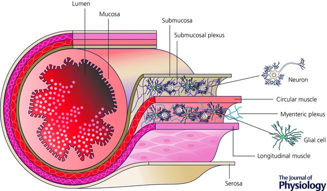
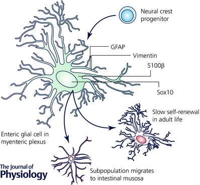
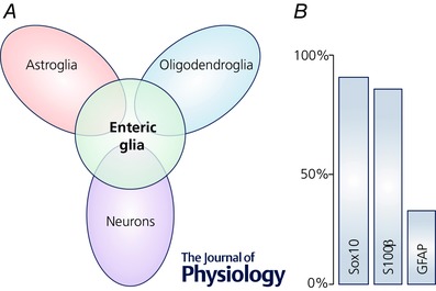
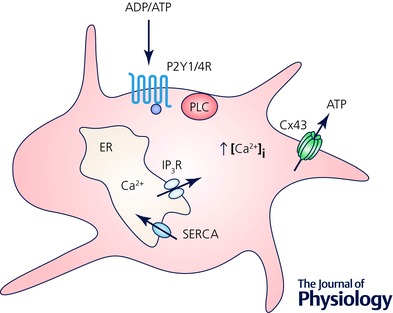
Glia (from Greek γλοία meaning 'glue') pertains to non-neuronal cells in the central (CNS) and peripheral nervous system (PNS) that nourish neurons and maintain homeostasis. In addition, glia are now increasingly appreciated as active regulators of numerous physiological processes initially considered exclusively under neuronal regulation. For instance, enteric glia, a collection of glial cells residing within the walls of the intestinal tract, regulate intestinal motility, a well-characterized reflex controlled by enteric neurons. Enteric glia also interact with various non-neuronal cell types in the gut wall such as enterocytes, enteroendocrine and immune cells and are therefore emerging as important local regulators of diverse gut functions. The intricate molecular mechanisms that govern glia-mediated regulation are beginning to be discovered, but much remains unknown about the functions of enteric glia in health and disease. Here we present a current view of the enteric glia and their regulatory roles in gastrointestinal (GI) (patho)physiology; from GI motility and epithelial barrier function to enteric neuroinflammation.


Figures

Figure 1
Figure 1. Schematic depiction of the intestine showing the general arrangement of the enteric nervous system in the gut wall
Enteric neurons and glia are housed within the submucosal and myenteric plexuses. Neural programmes in the submucosal plexus regulate fluid exchange across the intestinal mucosa and neural programmes in the myenteric plexus coordinate the contractile activity of the intestine. Image courtesy of David E. Fried.
Figure 2
Figure 2. Enteric glial cells derive from neural crest precursors and mature into neuroglia in the enteric nervous system
Enteric glia within the myenteric plexus are slowly replaced under physiological conditions (Joseph et al. 2011; Laranjeira et al. 2011) and are responsible for generating glia that migrate to the intestinal mucosa (Kabouridis et al. 2015).
Figure 3
Figure 3. Gene expression in enteric glia
A, transcriptional profile of enteric glia compared with the profiles of neurons and glia from the CNS (Rao et al. 2015). Not drawn to scale. B, expression of common markers for enteric glia estimated from Boesmans et al. (2015); co‐localization among the glial markers omitted for clarity.
Figure 4
Figure 4. Enteric glia actively participate in purinergic neuron–glia signalling
ADP and ATP bind to G‐protein coupled purinergic receptors P2Y1R and P2Y4R, respectively, and activate phospholipase C (PLC) and subsequent production of inositol 1,4,5‐trisphosphate (IP3). This consequently activates IP3 receptors (IP3R) inducing the release of Ca2+ from endoplasmic reticulum (ER). Increase in the intracellular Ca2+ concentration [Ca2+]i induces ATP release through Cx43 hemichannels. Sarco/endoplasmic reticulum Ca2+‐ATPase (SERCA) pumps Ca2+ ions back into the ER. Based from original research on enteric glial cells (Kimball & Mulholland 1996; Zhang et al. 2003; McClain et al. 2014). Not drawn to scale.
Figure 5
Figure 5. Enteric glia actively regulate gut motility
Inhibition (A–C) or activation (D–F) of glial calcium (Ca2+) signalling (A and D) results in reduction or stimulation of the gut motor reflex assessed by smooth muscle tension recordings (B and E), respectively, and corresponds to changes in the distal colon motility tested in vivo (C and F). A–C, experiments from tamoxifen‐induced glia‐specific knock out (igKO) of connexin 43 (Cx43) mice (Cx43‐igKO) and the tamoxifen‐treated background strains (Backgr.); figures obtained from McClain et al. (2014). A, neuron‐specific stimulation activates Ca2+ responses in enteric glia and Cx43 is required for the propagation of the glial Ca2+ response (see original work for details). B, electrical field stimulation (EFS) elicits muscle contractions and the contraction force is reduced in the Cx43‐igKO mice. C, selective reduction of the Ca2+ response in the enteric glia reduces distal colon motility in vivo. D–F, experiments from GFAP::hM3Dq transgenic mice, where glial fibrillary acidic protein (GFAP) promoter drives expression of an engineered Gq‐coupled human M3 muscarinic receptor (hM3Dq) and their wild‐type (WT) littermates; figures obtained from McClain et al. (2015). D, enteric glia expressing hM3Dq respond to clozapine N‐oxide (CNO) with an increase in cytosolic Ca2+ and subsequently affect neurally controlled gut reflexes. E, glia‐specific stimulation with CNO evoked response in GFAP::hM3Dq mice similar to stimuli with bethanechol (BCH) and EFS that directly activate smooth muscle and enteric neurons, respectively. Note that CNO effect was blocked by tetrodotoxin (TTX) indicating that glia‐specific effects are mediated via enteric neurons. Also, CNO stimulation evoked no response in WT littermates (see original work). F, selective activation of glial Ca2+ signalling enhances in vivo motility of the distal colon.
Figure 6
Figure 6. Enteric glia as active players in the peristaltic reflex
The accepted circuitry of the peristaltic reflex involves the following chain of events: (1) mechanical or chemical stimuli in the gut lumen activate intrinsic primary afferent neurons (IPANs) residing in both plexi; (2) IPANs activate interneurons that project in both oral (ascending) and aboral (descending) directions; (3) ascending interneurons activate excitatory motorneurons that cause smooth muscle contraction by releasing acetylcholine (ACh) and neuropeptides while descending interneurons produce relaxation below the point of stimulation by activating inhibitory motorneurons that release nitric oxide (NO), purines and other inhibitory molecules (Kunze & Furness, 1999). Enteric glia cells (EGC) could interact with the circuit at multiple levels (see text for details), from the release of serotonin from enterochromafine cells (EC) to the direct interaction with the smooth muscle cells. Other abbreviations: MP, myenteric plexus; SMP, submucosal plexus. This schematic representation is not drawn to scale.
Figure 7
Figure 7. The role of enteric glia in inflammation – feed‐forward loop leading to increased cell death
Both ATP and nitric oxide (NO) released from enteric glia regulate normal gut physiology (see text for details). Infection‐induced TLR signalling increases iNOS expression via NF‐κB and results in increased release of NO, a molecule with an antimicrobial effect. Excessive NO release, either by the infection or by other inflammatory signals (omitted for clarity) can also damage the cells leading to a surge of purines and S100β. While S100β enhances NO release via the increased iNOS expression, purine signalling increases intracellular calcium concentration ([Ca2+]i) and increased ATP release via Cx43 hemichannels. Increased [Ca2+] can also lead to increased iNOS activity and expression via CaMKII and PKC, respectively. Both PKC and CaMKII were not directly investigated in enteric glia (light grey); our findings indirectly show that PKC does not play a role in enteric glia (dashed arrows). The main findings are summarized from Esposito et al. (2014), Turco et al. (2014) and Brown et al. (2015); see text for details. Abbreviations: CaMKII, Ca2+/calmodulin‐dependent protein kinase II; eNTPDase, ecto‐nucleoside triphosphate diphosphohydrolase; iNOS, inducible nitric oxide synthase; NF‐κB, nuclear factor kappa‐light‐chain‐enhancer of activated B cells; PKC, protein kinase C; PLC, phospholipase C; PPARα, peroxisome‐proliferator‐activated receptor‐α; S100β, S100 calcium‐binding protein β; TLR, toll‐like receptor.

References

    1. Abreu MT (2010). Toll‐like receptor signalling in the intestinal epithelium: how bacterial recognition shapes intestinal function. Nat Rev Immunol 10, 131–144. - PubMed
    1. Aube AC, Cabarrocas J, Bauer J, Philippe D, Aubert P, Doulay F, Liblau R, Galmiche JP & Neunlist M (2006). Changes in enteric neurone phenotype and intestinal functions in a transgenic mouse model of enteric glia disruption. Gut 55, 630–637. - PMC - PubMed
    1. Bach‐Ngohou K, Mahe MM, Aubert P, Abdo H, Boni S, Bourreille A, Denis MG, Lardeux B, Neunlist M & Masson D (2010). Enteric glia modulate epithelial cell proliferation and differentiation through 15‐deoxy‐12,14‐prostaglandin J2. J Physiol 588, 2533–2544. - PMC - PubMed
    1. Badizadegan K, Thomas AR, Nagy N, Ndishabandi D, Miller SA, Alessandrini A, Belkind‐Gerson J & Goldstein AM (2014). Presence of intramucosal neuroglial cells in normal and aganglionic human colon. Am J Physiol Gastrointest Liver Physiol 307, G1002–G1012. - PMC - PubMed
    1. Barajon I, Serrao G, Arnaboldi F, Opizzi E, Ripamonti G, Balsari A & Rumio C (2009). Toll‐like receptors 3, 4, and 7 are expressed in the enteric nervous system and dorsal root ganglia. J Histochem Cytochem 57, 1013–1023. - PMC - PubMed

Publication types

LinkOut - more resources