Randomized Controlled Trial
. 2017 Mar;25(3):876-886.
doi: 10.1007/s00167-016-4131-0. Epub 2016 Apr 22.

Knee joint distraction compared with high tibial osteotomy: a randomized controlled trial

Affiliations
Randomized Controlled Trial

Knee joint distraction compared with high tibial osteotomy: a randomized controlled trial

J A D van der Woude et al. Knee Surg Sports Traumatol Arthrosc. 2017 Mar.

Abstract

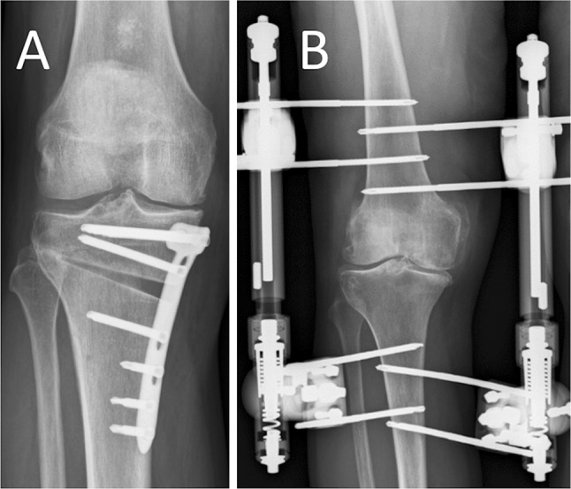
Purpose: Both, knee joint distraction as a relatively new approach and valgus-producing opening-wedge high tibial osteotomy (HTO), are knee-preserving treatments for knee osteoarthritis (OA). The efficacy of knee joint distraction compared to HTO has not been reported.

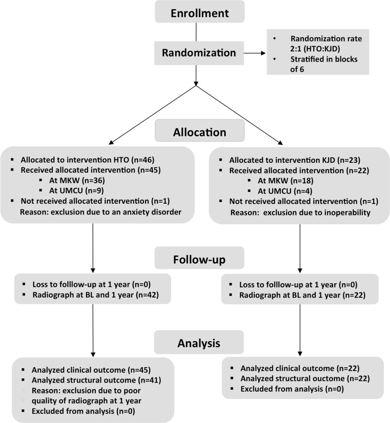
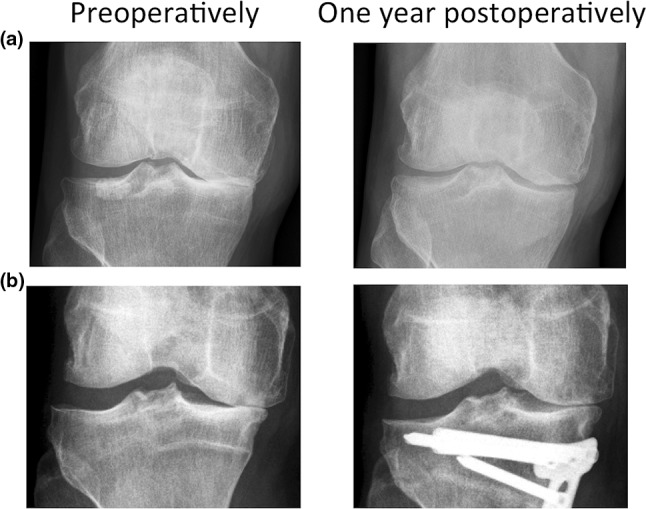
Methods: Sixty-nine patients with medial knee joint OA with a varus axis deviation of <10° were randomized to either knee joint distraction (n = 23) or HTO (n = 46). Questionnaires were assessed at baseline and 3, 6, and 12 months. Joint space width (JSW) as a surrogate measure for cartilage thickness was determined on standardized semi-flexed radiographs at baseline and 1-year follow-up.

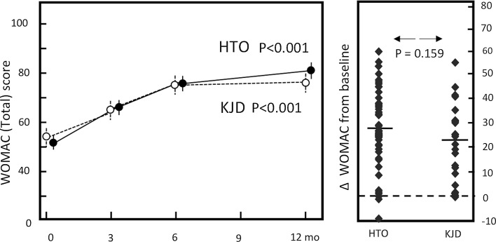
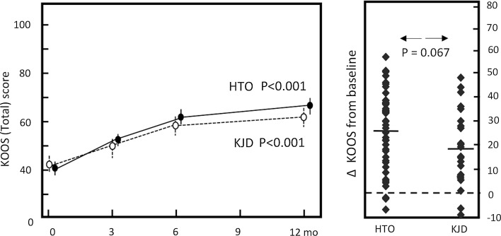
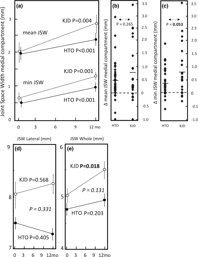
Results: All patient-reported outcome measures (PROMS) improved significantly over 1 year (at 1 year p < 0.02) in both groups. At 1 year, the HTO group showed slightly greater improvement in 4 of the 16 PROMS (p < 0.05). The minimum medial compartment JSW increased 0.8 ± 1.0 mm in the knee joint distraction group (p = 0.001) and 0.4 ± 0.5 mm in the HTO group (p < 0.001), with minimum JSW improvement in favour of knee joint distraction (p = 0.05). The lateral compartment showed a small increase in the knee joint distraction group and a small decrease in the HTO group, leading to a significant increase in mean JSW for knee joint distraction only (p < 0.02).

Conclusion: Cartilaginous repair activity, as indicated by JSW, and clinical outcome improvement occurred with both, knee joint distraction and HTO. These findings suggest that knee joint distraction may be an alternative therapy for medial compartmental OA with a limited mechanical leg malalignment.

Level of evidence: Randomized controlled trial, Level I.

Keywords: Cartilage repair; High tibial osteotomy; Joint distraction; Knee osteoarthritis.


Conflict of interest statement

None of the authors have a conflict of interest to declare.

Figures

Fig. 1
Fig. 1
Example of a post-operative radiograph, a patient treated with HTO, b patient treated with knee joint distraction
Fig. 2
Fig. 2
Flow chart including the numbers of excluded patients, as well allocation of the randomized treatment and the analysed patients per treatment arm. KJD knee joint distraction, MKW Maartenskliniek Woerden, UMCU University Medical Center Utrecht
Fig. 3
Fig. 3
WOMAC total. Dotted line represents the knee joint distraction group (n = 22), solid line represents the HTO group (n = 45). Mean values ± SEM are given. p values show statistical difference in values at 1-year follow-up compared to pre-treatment values. Mean change of WOMAC total (right): for both groups (average: dash) and for every individual patient (squares)
Fig. 4
Fig. 4
KOOS total. Dotted line represents the knee joint distraction group (n = 22), solid line represents the HTO group (n = 45). Mean values ± SEM are shown. p values show statistical difference in values at 1-year follow-up compared to pre-treatment values. Mean change of KOOS total score (right): for both groups (average: dash) and for every individual patient (squares)
Fig. 5
Fig. 5
a Mean quantitative radiograph analyses of the medial (affected) compartment of both treatment groups. The solid line represents HTO group (n = 41), and the dotted line represents the knee joint distraction group (n = 22). Mean values ± SEM are presented. p values show statistical difference in values at 1-year follow-up compared to pre-treatment values. b Mean change of mean JSW of medial compartment. For both groups (average: dash) and for every individual patient (squares). c Mean change of minimum JSW of medial compartment. For both groups (average: dash) and for every individual patient (squares). d Mean JSW of the lateral (least affected) compartment of both treatment groups. e Mean JSW of the whole joint of the both treatment groups. The p value in italic shows statistical difference in change over 1 year between the two treatment groups
Fig. 6
Fig. 6
a Radiograph preoperatively (left) and 1 year post-operatively (right) of a representative patient treated with knee joint distraction. b Radiograph preoperatively (left) and 1 year post-operatively (right) of a representative patient treated with HTO

References

    1. Abouheif MM, Nakamura M, Deie M, Adachi N, Nishimori M, Sera S, et al. Repair of a large osteochondral defect in the knee joint using autologous and artificial bone graft combined with motion preserving distraction arthroplasty: a case report. Arch Orthop Trauma Surg. 2010;130(2):231–236. doi: 10.1007/s00402-009-0998-2. - DOI - PubMed
    1. Aly TA, Hafez K, Amin O. Arthrodiatasis for management of knee osteoarthritis. Orthopedics. 2011;34(8):338–343. - PubMed
    1. Anagnostakos K, Mosser P, Kohn D. Infections after high tibial osteotomy. Knee Surg Sports Traumatol Arthrosc. 2013;21(1):161–169. doi: 10.1007/s00167-012-2084-5. - DOI - PubMed
    1. Baboolal TG, Mastbergen SC, Jones E, Calder SJ, Lafeber FP, McGonagle D. Synovial fluid hyaluronan mediates MSC attachment to cartilage, a potential novel mechanism contributing to cartilage repair in osteoarthritis using knee joint distraction. Ann Rheum Dis. 2015 - PMC - PubMed
    1. Birmingham TB, Giffin JR, Chesworth BM, Bryand DM, Litchfield RB, Willits K, et al. Medial opening wedge high tibial osteotomy: a prospective cohort study of gait, radiographic, and patient-reported outcomes. Arthritis Rheum. 2009;61(5):648–657. doi: 10.1002/art.24466. - DOI - PubMed

Publication types

LinkOut - more resources