Review
. 2016 Jun;7(3):365-71.
doi: 10.1007/s13244-016-0491-8. Epub 2016 Apr 23.

Multi-modal imaging of adhesive capsulitis of the shoulder

Affiliations
Review

Multi-modal imaging of adhesive capsulitis of the shoulder

Marcello Zappia et al. Insights Imaging. 2016 Jun.

Abstract

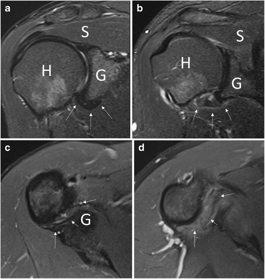
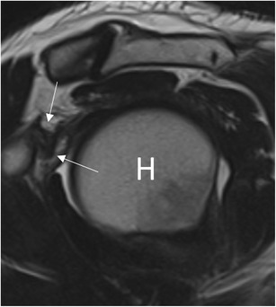
Adhesive capsulitis of the shoulder is a clinical condition characterized by progressive limitation of active and passive mobility of the glenohumeral joint, generally associated with high levels of pain. Although the diagnosis of adhesive capsulitis is based mainly on clinical examination, different imaging modalities including arthrography, ultrasound, magnetic resonance, and magnetic resonance arthrography may help to confirm the diagnosis, detecting a number of findings such as capsular and coracohumeral ligament thickening, poor capsular distension, extracapsular contrast leakage, and synovial hypertrophy and scar tissue formation at the rotator interval. Ultrasound can also be used to guide intra- and periarticular procedures for treating patients with adhesive capsulitis.

Key points: • Diagnosis of adhesive capsulitis is mainly based on clinical findings. • Imaging may be used to exclude articular or rotator cuff pathology. • Thickening of coracohumeral and inferior glenohumeral ligaments are common findings. • Rotator interval fat pad obliteration has 100 % specificity for adhesive capsulitis. • Ultrasound can be used to guide intra- and periarticular treatments.

Keywords: Adhesive capsulitis; Arthrography; Magnetic resonance; Shoulder; Ultrasound.


Figures

Fig. 1
Fig. 1
Conventional arthrography, anteroposterior view. (a) Normal distension of the axillary recess (black arrow) and the subscapular recess (thick arrow). (b) Reduced distension of the axillary recess (black arrow) and subscapular recess associated with medial leakage of contrast (white arrow) in a patient with adhesive capsulitis
Fig. 2
Fig. 2
Long-axis ultrasound scan of the proximal portion of the coracohumeral ligament (calipers) in a patient with adhesive capsulitis. The ligament is hypoechoic and thickened (1.8 mm). H humerus, arrow coracoid process
Fig. 3
Fig. 3
Axillary long-axis view of the inferior glenohumeral ligament with arm in abduction. (a) Thickening of the inferior capsular profile (calipers, 3.3 mm) in a shoulder affected by adhesive capsulitis. H humerus (b) In the contralateral shoulder, the capsule has normal thickness (calipers, 1.5 mm)
Fig. 4
Fig. 4
Evaluation of the rotator interval in a patient with adhesive capsulitis for 3 weeks. The power Doppler signal is clearly seen within hypoechoic scar tissue (asterisks). H humerus, arrow biceps tendon
Fig. 5
Fig. 5
Coronal oblique T2-weighted fat-saturated (a, b) and axial proton density fat-saturated (c, d) images. In a healthy subject (a, c), the capsular recess has normal signal intensity (arrows), while in a patient with adhesive capsulitis (b, d), clear signal hyperintensity can be seen (arrows). H humerus, G glenoid, S supraspinatus tendon
Fig. 6
Fig. 6
Coronal oblique proton density image in a patient with adhesive capsulitis. The axillary pouch (arrows) is thickened. G glenoid, H humerus
Fig. 7
Fig. 7
Sagittal oblique T2-weighted image in a patient with adhesive capsulitis. The coracohumeral ligament (arrows) is markedly thickened
Fig. 8
Fig. 8
MR arthrography, sagittal oblique T1-weighted image. In a patient with adhesive capsulitis for 15 weeks, the fat triangle (arrowheads) signal is considerably reduced
Fig. 9
Fig. 9
MR arthrography, coronal oblique T1-weighted fat-saturated image. (a) In a healthy subject, the axillary pouch is normally distensible (arrow). H humerus, G glenoid. (b). In a patient with adhesive capsulitis, the axillary pouch is contracted and poorly distended (arrow)
Fig. 10
Fig. 10
MR arthrography, (a) coronal oblique and (b) axial T1-weighted fat-saturated image in a patient with adhesive capsulitis. Leakage of contrast agent can be seen on the anterior inferior margin of the scapula (arrow). H humerus, G glenoid

References

    1. Codman EA (1934) Rupture of the supraspinatus tendon and other lesions in or about the subacromial bursa. In: The shoulder. Privately printed, Boston. pp 216-224
    1. Mengiardi B, Pfirrmann CW, Gerber C, Hodler J, Zanetti M. Frozen shoulder: MR arthrographic findings. Radiology. 2004;233(2):486–492. doi: 10.1148/radiol.2332031219. - DOI - PubMed
    1. Neviaser JS. Adhesive capsulitis of the shoulder (the frozen shoulder) Med Times. 1962;90:783–780. - PubMed
    1. Harris G, Bou-Haidar P, Harris C. Adhesive capsulitis: review of imaging and treatment. J Med Imaging Radiat Oncol. 2013;57(6):633–643. doi: 10.1111/1754-9485.12111. - DOI - PubMed
    1. Sung CM, Jung TS, Park HB (2014) Are serum lipids involved in primary frozen shoulder? A case-control study. J Bone Joint Surg Am 96(21):1828–18s33 - PubMed