

Liver CT for vascular mapping during radioembolisation workup: comparison of an early and late arterial phase protocol
- PMID: 27108297
- PMCID: PMC5127855
- DOI: 10.1007/s00330-016-4343-1
Liver CT for vascular mapping during radioembolisation workup: comparison of an early and late arterial phase protocol
Abstract
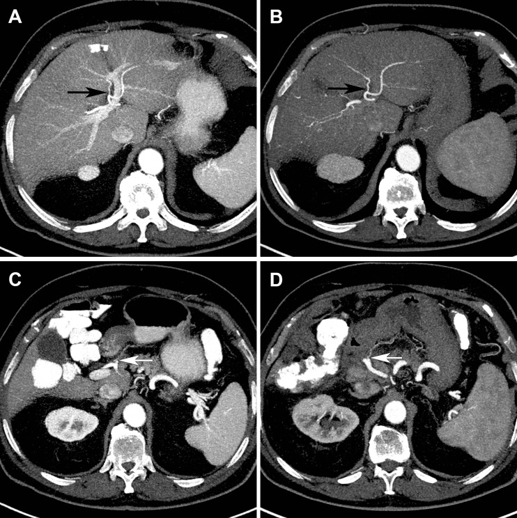
Objectives: To compare right gastric (RGA) and segment 4 artery (A4) origin detection rates during radioembolisation workup between early and late arterial phase liver CT protocols.
Methods: 100 consecutive patients who underwent liver CT between May 2012-January 2015 with early or late arterial phase protocol (n = 50 each, 10- vs. 20-s post-threshold delay) were included. RGA/A4 origin detection rates, assessed by two raters, and contrast-to-noise ratio (CNR) of the hepatic artery relative to the portal vein were compared between the protocols.
Results: The first-second rater scored the RGA origin as visible in 58-65 % (specific proportion of agreement 82 %, κ = 0.62); A4 origin in 96-89 % (94 %, κ = 0.54). Thirty-six percent of RGA origins not detectable by DSA were identified on CT. Origin detection rates were not significantly different for early/late arterial phases. Mean CNR was higher in the early arterial phase protocol (1.7 vs. 1.2, p < 0.001).
Conclusion: A 10-s delay arterial phase CT protocol does not significantly improve detection of small intra- and extrahepatic branches. RGA origin detection requires further optimization, whereas A4/MHA origin detection is adequate, with good inter-rater reproducibility. CT remains important for preprocedural planning, because it may reveal arterial anatomy not discernible on DSA.
Key points: • An early arterial phase does not significantly improve RGA and A4/MHA origin detection. • RGA origin detection (58-65 %) on CT is still suboptimal. • 36 % of RGA origins undetectable on DSA can be identified on CT. • A4/MHA origin detection (89-96 %) on CT is excellent. • Inter-rater reproducibility is good for RGA and A4/MHA origin detection on CT.
Keywords: Acquisition protocol; Arterial phase; Liver CT; Radioembolisation; SIRT.
Figures
References
-
- van den Hoven AF, van Leeuwen MS, Lam MGEH, van den Bosch MAAJ (2014) Hepatic arterial configuration in relation to the segmental anatomy of the liver; observations on MDCT and DSA relevant to radioembolization treatment. Cardiovasc Intervent Radiol 1–12 - PubMed
-
- Wicherts DA, de Haas RJ, van Kessel CS, et al. Incremental value of arterial and equilibrium phase compared to hepatic venous phase CT in the preoperative staging of colorectal liver metastases: an evaluation with different reference standards. Eur J Radiol. 2011;77:305–311. doi: 10.1016/j.ejrad.2009.07.026. - DOI - PubMed
Publication types
MeSH terms
LinkOut - more resources
Full Text Sources
Other Literature Sources
Medical
