. 2016 Apr;18(4):199-212.
doi: 10.1016/j.neo.2016.02.005. Epub 2016 Mar 22.

A Potent Derivative of Indolizino[6,7-b]Indole for Treatment of Human Non-Small Cell Lung Cancer Cells

Affiliations

A Potent Derivative of Indolizino[6,7-b]Indole for Treatment of Human Non-Small Cell Lung Cancer Cells

Chi-Wei Chen et al. Neoplasia. 2016 Apr.

Erratum in

Abstract

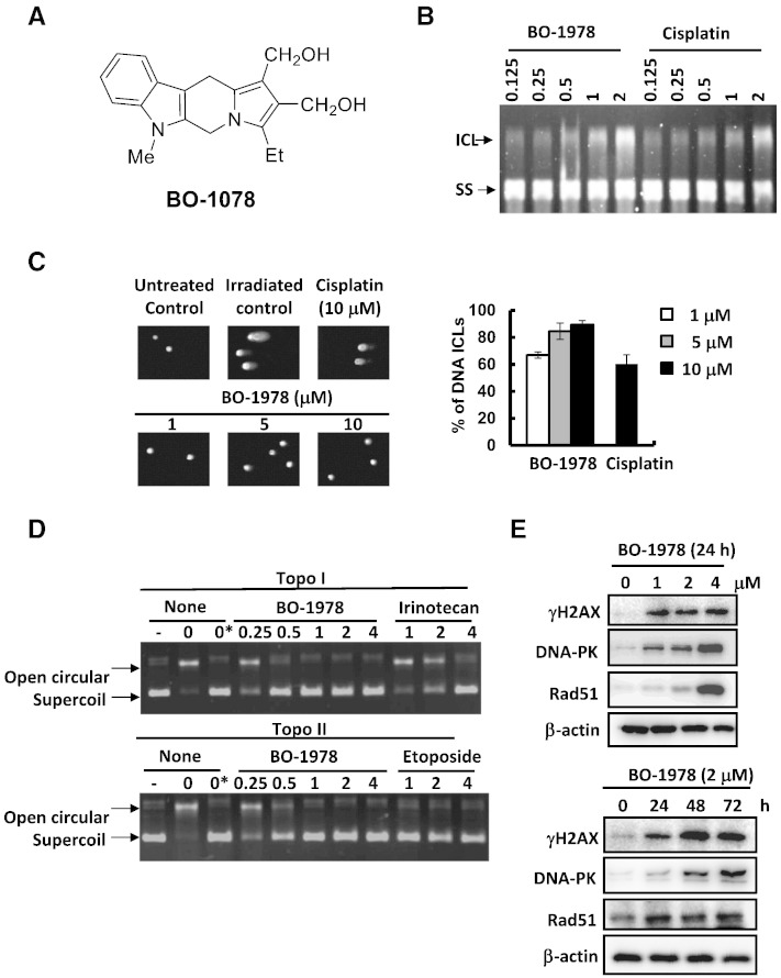
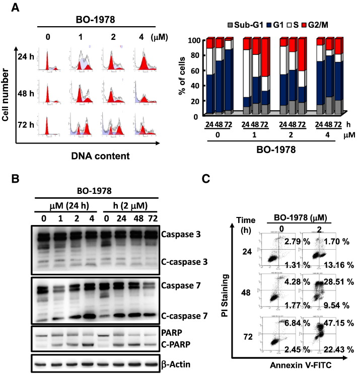
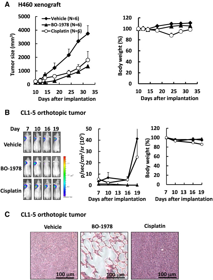
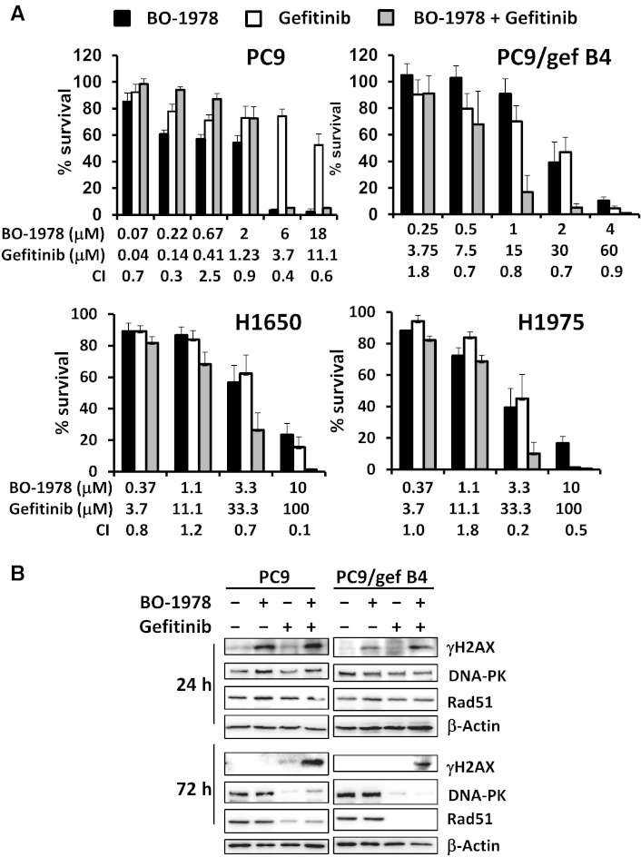
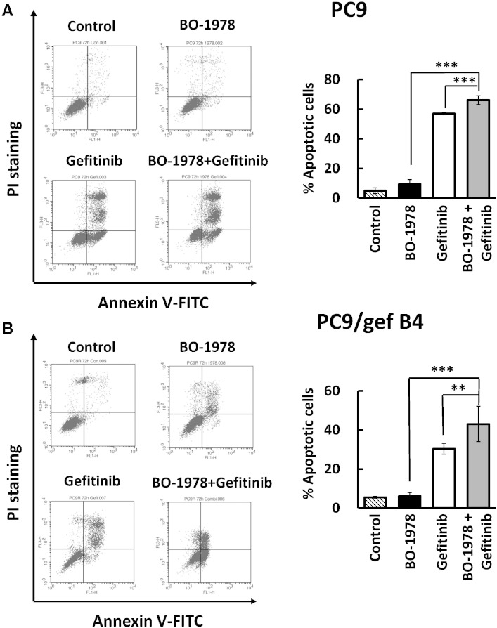
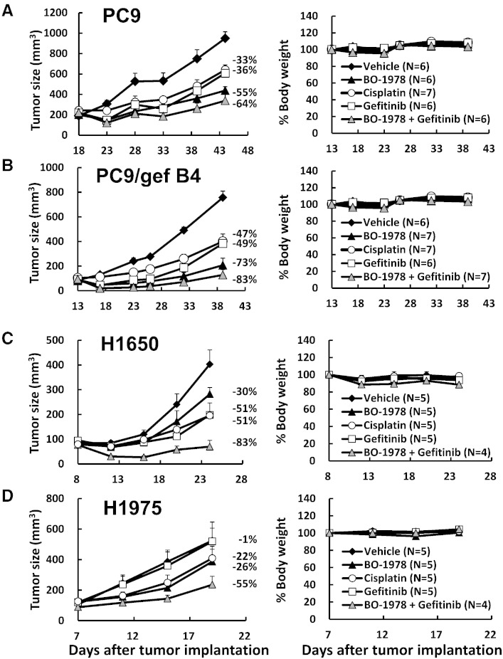
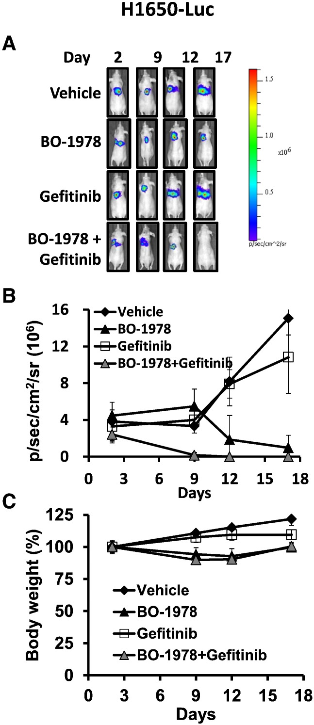
The therapeutic effect in non-small cell lung cancer (NSCLC) patients is limited because of intrinsic and acquired resistance. Thus, an unmet need exists for the development of new drugs to improve the therapeutic efficacy in NSCLC patients. In this study, the novel small molecule indolizino[6,7-b]indole derivative BO-1978 was selected to evaluate its therapeutic effects on NSCLC and its preclinical toxicity in animal models. An in vitro cytotoxicity assay revealed that BO-1978 significantly suppressed the growth of various NSCLC cell lines with or without mutations in epidermal growth factor receptor (EGFR). Mechanistically, we demonstrated that BO-1978 exhibited multiple modes of action, including inhibition of topoisomerase I/II and induction of DNA cross-linking. Treatment of NSCLC cells with BO-1978 caused DNA damage, disturbed cell cycle progression, and triggered apoptotic cell death. Furthermore, BO-1978 significantly suppressed the growth of EGFR wild-type and mutant NSCLC tumors in xenograft tumor and orthotopic lung tumor models with negligible body weight loss. The combination of BO-1978 with gefitinib further suppressed EGFR mutant NSCLC cell growth in xenograft tumor and orthotopic lung tumor models. Preclinical toxicity studies showed that BO-1978 administration did not cause apparent toxicity in mice. Based on its significant therapeutic efficacy and low drug toxicity, BO-1978 is a potential therapeutic agent for treatment of NSCLC.


Figures

Figure 1
Figure 1
DNA damage induced by BO-1978. (A) Chemical structure of BO-1978. (B) Formation of DNA cross-linking by BO-1978. Plasmid DNA was treated with various concentrations (0.125 to 2 μM) of BO-1978 or cisplatin for 2 hours at 37°C. The ICL and single-strand DNA was electrophoretically separated under alkaline conditions, as described in the Materials and Methods section. (C) Induction of DNA ICLs in H460 cells by BO-1978. H460 cells were treated with various concentrations (1, 5, and 10 μM) of BO-1978 or 10 μM cisplatin for 2 hours. DNA ICLs formed in H460 cells were identified using a modified comet assay. (D) Inhibition of intracellular topo I (upper panel) and II (lower panel) activities in H460 cells. The nuclear extracts of BO-1978–treated H460 cells (0.25 to 4 μM for 72 hours) were prepared and incubated with pEGFP-N1 plasmid DNA and specific topo I or topo II buffer for 30 minutes at 37°C as described in the Materials and Methods section. The relaxation of plasmid DNA was analyzed by electrophoresis. Irinotecan and etoposide served as positive controls for topo I and topo II, respectively. (E) The DNA damage response of H460 cells to BO-1978. H460 cells were treated with BO-1978 at the concentrations of 1 to 4 μM for 24 hours or 2 μM BO-1978 for various time periods (24 to 72 hours). γH2AX (Ser139), DNA-PK, and Rad51 protein expression levels were determined by western blotting.
Figure 2
Figure 2
Interference with cell cycle progression and induction of apoptosis by BO-1978 in H460 cells. (A) Cell cycle interference by BO-1978. H460 cells were treated with various concentrations of BO-1978 (1 to 4 μM) for various time periods (24 to 72 hours). The distribution of cell cycle phases was analyzed using a flow cytometer and ModFit LT 3.0 software (Verity Software House, Topsham, ME). (B) Activation of the apoptotic pathway by BO-1978. The appearance of cleaved caspase-3 and -7 and PARP in BO-1978–treated H460 cells was determined by Western blotting. (C) Induction of apoptosis by BO-1978. The apoptotic cells induced by BO-1978 were determined using an Annexin V–FITC apoptosis detection kit, as described in the Materials and Methods section.
Figure 3
Figure 3
The therapeutic efficacy of BO-1978 against EGFR wild-type NSCLC cells. (A) Suppression of H460 xenografts by BO-1978. H460 cells (3 × 10⁶) were subcutaneously implanted in nude mice. When the tumor size reached approximately 100 mm3, the tumor-bearing mice were intravenously treated with vehicle, BO-1978 (40 mg/kg, daily for 5 consecutive days), or cisplatin (6 mg/kg, three times every 4 days). The tumor size and body weight were measured at the times indicated. (B) Suppression of orthotopically implanted CL1-5/GFP-Luciferase cells by BO-1978. CL1-5/GFP-Luciferase cells (5 × 10⁶) were orthotopically implanted in the lungs of nude mice. The tumor-bearing mice were treated with vehicle, BO-1978, or cisplatin, as described above. (Left) The representative images of mice implanted with CL1-5/GFP-Luciferase cells and treated with drugs. (Middle and right) The quantitative signals of luciferase (p/s per cm2 per sr) and the relative body weights of mice treated with vehicle, BO-1978, or cisplatin, respectively. p, photon; sr, steradian. (C) No tumor formation in the lungs of orthotopically implanted mice treated with BO-1978. On day 19, the lungs were harvested from mice treated with vehicle, BO-1978, or cisplatin; histopathologically sectioned; and stained with hematoxylin and eosin.
Figure 4
Figure 4
Enhancement of BO-1978–induced toxic effects in EGFR mutant NSCLC cells upon gefitinib treatment. (A) Synergistic suppression of cell growth by combination treatment of EGFR mutant NSCLC with BO-1978 and gefitinib. Logarithmically growing PC9, PC9/gef B4, H1650, and H1975 cells were treated with BO-1978, gefitinib, or the combination for 72 hours. The cell growth was determined using an alamarBlue assay, as described in the Materials and Methods section. (B) Increased DNA damage marker (γH2AX) expression and suppression of DNA repair proteins (DNA-PK and Rad51) by gefitinib. PC9 and PC9/gef B4 cells were treated with BO-1978, gefitinib, or the combination for 24 and 72 hours. At the end of treatment, the cells were harvested, and γH2AX, DNA-PK, and Rad51 expression levels were analyzed by Western blotting.
Figure 5
Figure 5
Increasing BO-1978–induced apoptotic cells with gefitinib treatment in PC9 (A) and PC9/gef B4 cells (B). Logarithmically growing PC9 and PC9/gef B4 cells were treated with BO-1978 (2 μM), gefitinib (0.4 μM for PC9 cells and 30 μM for PC9/gef B4 cells), or the combination for 72 hours. At the end of treatment, the cells were harvested and subjected to analysis of apoptotic cells using an Annexin V–FITC apoptosis detection kit. The data on the right are the means and SD of three independent experiments. ** and ***, P < .01 and .001 according to Student’s t test.
Figure 6
Figure 6
Antitumor activity of BO-1978 in EGFR mutant NSCLC xenografts. Aliquots of 5 × 10⁶ PC9 (A), PC9/gef B4 (B), H1650 (C), and H1975 (D) cells were subcutaneously implanted in nude mice. When tumor sizes reached approximately 100 mm3, the mice were treated with vehicle, BO-1978 (40 mg/kg, daily for 5 consecutive days), gefitinib (10 mg/kg, daily for 5 consecutive days), BO-1978 + gefitinib, or cisplatin (6 mg/kg, three times every 4 days). The tumor volumes and body weights were monitored at the indicated times.
Figure 7
Figure 7
Suppression of orthotopic growth of H1650-Luc cells by BO-1978 alone or in combination with gefitinib. H1650-Luc cells (2 × 10⁶) were intrathoracically implanted in the left lobes of nude mice. On day 2, the mice were treated with BO-1978 alone or in combination with gefitinib, as described in Figure 6. Tumor growth was monitored by IVIS. (A) Representative images of tumor growth; (B) averaged photon intensity; and (C) body weight.

References

    1. Edwards BK, Noone AM, Mariotto AB, Simard EP, Boscoe FP, Henley SJ, Jemal A, Cho H, Anderson RN, Kohler BA. Annual report to the nation on the status of cancer, 1975-2010, featuring prevalence of comorbidity and impact on survival among persons with lung, colorectal, breast, or prostate cancer. Cancer. 2014;120:1290–1314. - PMC - PubMed
    1. Ettinger DS, Akerley W, Borghaei H, Chang AC, Cheney RT, Chirieac LR, D'Amico TA, Demmy TL, Govindan R, Grannis FW., Jr. Non–small cell lung cancer, version 2.2013. J Natl Compr Canc Netw. 2013;11:645–653. [quiz 653] - PubMed
    1. Cataldo VD, Gibbons DL, Perez-Soler R, Quintas-Cardama A. Treatment of non–small-cell lung cancer with erlotinib or gefitinib. N Engl J Med. 2011;364:947–955. - PubMed
    1. Clegg A, Scott DA, Hewitson P, Sidhu M, Waugh N. Clinical and cost effectiveness of paclitaxel, docetaxel, gemcitabine, and vinorelbine in non–small cell lung cancer: a systematic review. Thorax. 2002;57:20–28. - PMC - PubMed
    1. Lwin Z, Riess JW, Gandara D. The continuing role of chemotherapy for advanced non–small cell lung cancer in the targeted therapy era. J Thorac Dis. 2013;5:S556–S564. - PMC - PubMed