. 2016 Jun;35(6):3143-50.
doi: 10.3892/or.2016.4732. Epub 2016 Apr 4.

Expression of tropomyosin 2 gene isoforms in human breast cancer cell lines

Affiliations

Expression of tropomyosin 2 gene isoforms in human breast cancer cell lines

Syamalima Dube et al. Oncol Rep. 2016 Jun.

Abstract

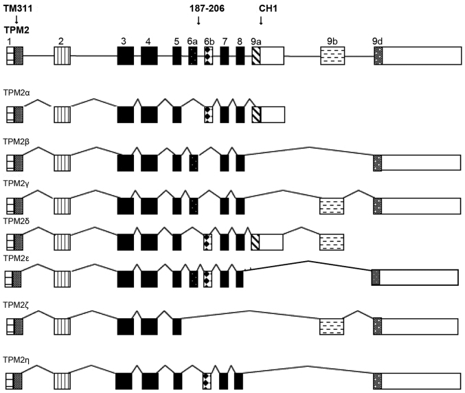
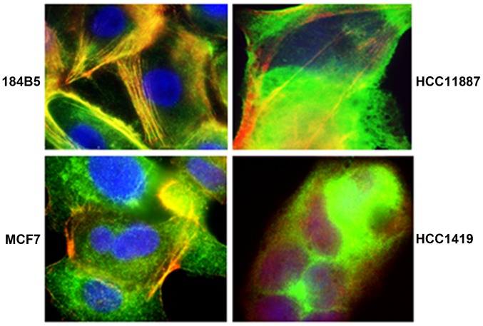
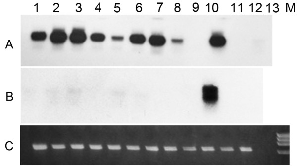
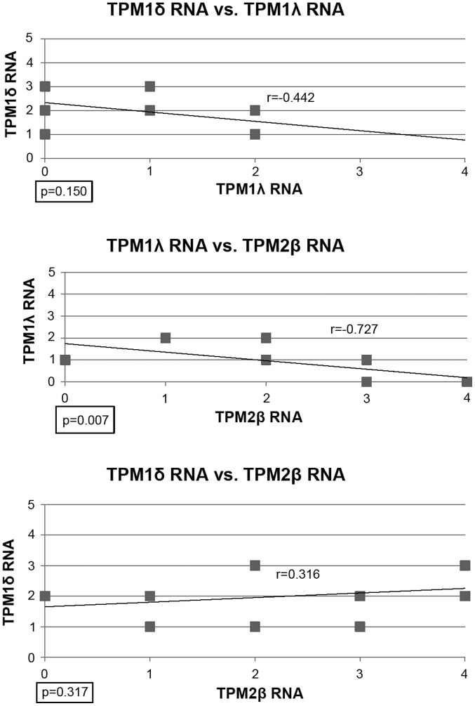
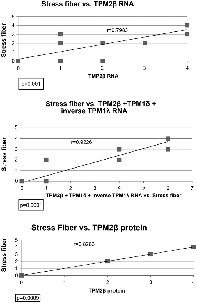
In humans, four tropomyosin genes (TPM1, TPM2, TPM3, and TPM4) are known to produce a multitude of isoforms via alternate splicing and/or using alternate promoters. Expression of tropomyosin has been shown to be modulated at both the transcription and the translational levels. Tropomyosins are known to make up some of the stress fibers of human epithelial cells and differences in their expression has been demonstrated in malignant breast epithelial cell lines compared to 'normal' breast cell lines. We have recently reported the expression of four novel TPM1 isoforms (TPM1λ, TPM1µ, TPM1ν, and TPM1ξ) from human malignant tumor breast cell lines that are not expressed in adult and fetal cardiac tissue. Also, we evaluated their expression in relation to the stress fiber formation. In this study, nine malignant breast epithelial cell lines and three 'normal' breast cell lines were examined for stress fiber formation and expression of tropomyosin 2 (TPM2) isoform-specific RNAs and proteins. Stress fiber formation was assessed by immunofluorescence using Leica AF6000 Deconvolution microscope. Stress fiber formation was strong (++++) in the 'normal' cell lines and varied among the malignant cell lines (negative to +++). No new TPM2 gene RNA isoforms were identified, and TPM2β was the most frequently expressed TPM2 RNA and protein isoform. Stress fiber formation positively correlated with TPM2β RNA or protein expression at high, statistically significant degrees. Previously, we had shown that TPM1δ and TPM1λ positively and inversely, respectively, correlated with stress fiber formation. The most powerful predictor of stress fiber formation was the combination of TPM2β RNA, TPM1δ RNA, and the inverse of TPM1λ RNA expression. Our results suggest that the increased expression of TPM1λ and the decreased expression of TPM1δ RNA and TPM2β may lead to decreased stress fiber formation and malignant transformation in human breast epithelial cells.


Figures

Figure 1
Figure 1
The various exons (boxes) and introns (lines) of the human tropomyosin 2 gene and its various RNA isoforms identified to date. The exons identified by the antibodies TM311, which goes against all four TPM genes, 187–200, which is TPM2-specific, and CH1, which goes against the TPM1, 2 and 3 genes, are identified. TPM1E2α is not shown because it goes against only the TPM1 gene. Those exons that are translated as peptides are indicated by solid or hatched, markings, while those that are not, are left blank.
Figure 2
Figure 2
Merged photograph of one 'normal' (184B5) and three malignant (HCC1187, MCF7, and HCC1419) human breast cell lines after staining with TM311, phalloidin and DAPI. The stress fibers are the linear lines that contain both actin and tropomyosin resulting in a variety of colors depending on the background, while the nuclei are stained blue with DAPI. The cell lines were given the following stress fiber scores: 184B5 ++++, HCC1187 ++, and MCF7 negative, and HCC1419 negative.
Figure 3
Figure 3
Expression of TPM2α and TPM2β transcripts in various malignant and non-malignant human breast tumor cell lines. A: Southern hybridization of TPM2 cDNA amplified with TPM2, Exon 1a(+) and TPM2, exon 9d(−) primers, and probed with radio-labeled TPM2 exon 5(+) oligonucleotide probe. B: Southern hybridization of cDNA amplified with TPM2, exon 1a(+) and TPM2, exon 9a(−) and probed with radio labeled TPM2 exon 5(+) oligonucleotide probe. C: Ethidium bromide-stained RT-PCR amplified DNA of human GAPDH. Lane 1, MCF7; lane 2, MCF-10A; lane 3, HCC-1143; lane 4, HCC-1143 BL; lane 5, MDA-MB-453; lane 6, MDA-MB-468; lane 7, HCC-1806; lane 8, HCC-1187; lane 9, BT-474; lane 10, MDA-MB-157; lane 11, HCC-1419; lane 12, HCC-1419 BL; lane 13, primer control.
Figure 4
Figure 4
Regression analysis of TPM1δ RNA vs. TPM1λ RNA; TPM1δ RNA vs. TPM2β RNA or TPM1λ RNA vs. TPM2β RNA in the 12 human breast cell lines. Each box represents one, or more than one cell line.
Figure 5
Figure 5
Regression analyses of stress fiber formation vs. expression of TPM2β RNA, TPM2β protein, or TPM2β RNA plus TPM1δ RNA plus the inverse of TPM1λ RNA in the 12 human breast cell lines. Each box represents one, or more than one, cell line. Both TPM2β RNA and protein expression had high positive correlations with stress fiber formation (both p=0.001). The highest correlation with stress fiber formation (r=0.92) was observed when TPM2 RNA, TPM1δ RNA and the inverse of TPM1λ RNA were combined (p<0.001).

References

    1. Gunning P, O'Neill G, Hardeman E. Tropomyosin-based regulation of the actin cytoskeleton in time and space. Physiol Rev. 2008;88:1–35. doi: 10.1152/physrev.00001.2007. - DOI - PubMed
    1. Lees-Miller JP, Helfman DM. The molecular basis for tropomyosin isoform diversity. BioEssays. 1991;13:429–437. doi: 10.1002/bies.950130902. - DOI - PubMed
    1. Perry SV. Vertebrate tropomyosin: Distribution, properties and function. J Muscle Res Cell Motil. 2001;22:5–49. doi: 10.1023/A:1010303732441. - DOI - PubMed
    1. Pieples K, Wieczorek DF. Tropomyosin 3 increases striated muscle isoform diversity. Biochemistry. 2000;39:8291–8297. doi: 10.1021/bi000047x. - DOI - PubMed
    1. Pittenger MF, Kazzaz JA, Helfman DM. Functional properties of non-muscle tropomyosin isoforms. Curr Opin Cell Biol. 1994;6:96–104. doi: 10.1016/0955-0674(94)90122-8. - DOI - PubMed