Case Reports
. 2016 May 5;98(5):971-980.
doi: 10.1016/j.ajhg.2016.03.004. Epub 2016 Apr 21.

Recessive and Dominant De Novo ITPR1 Mutations Cause Gillespie Syndrome

Affiliations
Case Reports

Recessive and Dominant De Novo ITPR1 Mutations Cause Gillespie Syndrome

Sylvie Gerber et al. Am J Hum Genet. .

Abstract

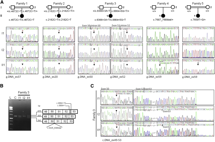
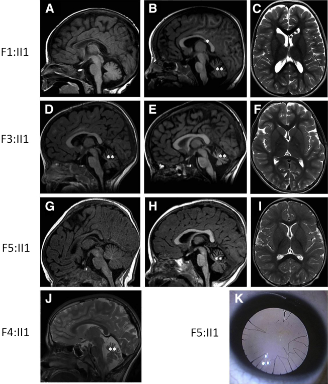
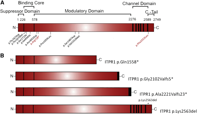
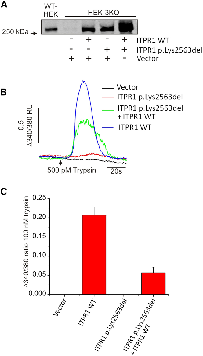
Gillespie syndrome (GS) is a rare variant form of aniridia characterized by non-progressive cerebellar ataxia, intellectual disability, and iris hypoplasia. Unlike the more common dominant and sporadic forms of aniridia, there has been no significant association with PAX6 mutations in individuals with GS and the mode of inheritance of the disease had long been regarded as uncertain. Using a combination of trio-based whole-exome sequencing and Sanger sequencing in five simplex GS-affected families, we found homozygous or compound heterozygous truncating mutations (c.4672C>T [p.Gln1558(∗)], c.2182C>T [p.Arg728(∗)], c.6366+3A>T [p.Gly2102Valfs5(∗)], and c.6664+5G>T [p.Ala2221Valfs23(∗)]) and de novo heterozygous mutations (c.7687_7689del [p.Lys2563del] and c.7659T>G [p.Phe2553Leu]) in the inositol 1,4,5-trisphosphate receptor type 1 gene (ITPR1). ITPR1 encodes one of the three members of the IP3-receptors family that form Ca(2+) release channels localized predominantly in membranes of endoplasmic reticulum Ca(2+) stores. The truncation mutants, which encompass the IP3-binding domain and varying lengths of the modulatory domain, did not form functional channels when produced in a heterologous cell system. Furthermore, ITPR1 p.Lys2563del mutant did not form IP3-induced Ca(2+) channels but exerted a negative effect when co-produced with wild-type ITPR1 channel activity. In total, these results demonstrate biallelic and monoallelic ITPR1 mutations as the underlying genetic defects for Gillespie syndrome, further extending the spectrum of ITPR1-related diseases.


Figures

Figure 1
Figure 1
Recessive and Dominant De Novo ITPR1 Mutations in Families with GS (A) Pedigree of the families and chromatograms showing the mutations at the genomic level. In families F1, F2, and F3, the affected individual is homozygous or compound heterozygous for parental mutations whereas in families F4 and F5, the affected individual is single heterozygous for a mutation absent in parental DNA, suggesting a de novo event. (B) Agarose gel separation of PCR products obtained from reverse-transcribed leucocyte mRNAs from individuals of family F3 and a control subject using the following primers: forward (exon 49), 5′-caatgcctcgaagttgctc-3′ and reverse (exon 53) 5′-cgtgggttgtaacccgact-3′. A Formamide gel-loading buffer was used to avoid formation of heteroduplexes. The 605 bp wild-type fragment in the control is seen in all three members of the family. The presence of this band in II1 demonstrates that both the c.6366+3A>T and c.6664+5G>T allow the production of a wild-type transcript (chromatogram not shown). The mutant mRNA transcribed from the c.6366+3A>T and c.6664+5G>T alleles are seen as additional 543 bp and 615 bp fragments in I1 and II1 and I2 and II1, respectively. (C) Sanger sequencing of additional 543 and 615 bp fragments amplified from reverse-transcribed mRNAs from individuals of family F3 is consistent with the activation of cryptic splice sites in exon 50 and intron 52, respectively.
Figure 2
Figure 2
Brain MRI and Eye Findings in Individuals with Mutations in ITPR1 (A–J) MRIs were performed at 1.5 months and 4.5 years, 2.5 and 8 years, 3.5 months and 1.5 years, and 8 years of age in individuals F1:II1 (A–C), F3:II1 (D–F), F5:II1 (G–I), and F4:II1 (J) by means of the following modalities: sagittal T1 (A, B, D, E, G, H) or T2 (J) and axial T2 (C, F, I). In the first months of life, sagittal T1 images are unremarkable (A, G). The cerebellar atrophy appears with age as shown on sagittal T1 or T2 images by the moderate atrophy at age 1.5 years and 2.5 years (∗∗) (D, H) and the marked cerebellar atrophy at 4.5 and 8 years (∗∗) (B, D, J). In addition to cerebellar atrophy, individual F1:II1 presents with thin corpus callosum () (B) and discrete ventricular dilatation (∗∗∗) (C). (K) Photograph of an eye of individual F5:II1 showing typical partial iris hypoplasia with iris strands extending onto the anterior lens surface.
Figure 3
Figure 3
Schematic Diagrams Depicting Full-Length ITPR1 and ITPR1 Mutation Associated with Human Diseases (A) The panel displays the functional domains of ITPR1. Shown are point mutations associated with spinocerebellar ataxia or ataxic cerebellar palsy (blue) or Gillespie syndrome not investigated in this study (red). (B) The lower four panels show the truncation mutants and p.Lys2563del mutant expressed in heterologous cell systems for functional analysis. The black boxes of the ITPR1 p.Gly2102Valfs5 and ITPR1 p.Ala2221Valfs23 mutants depict the VGSA and VGFQRISNNTERHSSGISQTWA amino acid sequences introduced by the c.6366+3A>T and c.6664+5G>T, respectively.
Figure 4
Figure 4
Production of the ITPR1 p.Lys2563del Mutant in HEK-3KO and Evaluation of Its Effect on the Formation of ITPR1 Channels (A) HEK-3KO cells were transfected as indicated. Lysates were prepared from transfected cells, quantified and equivalent amounts of proteins were separated on 4% SDS-PAGE and processed in immunoblots using a rabbit polyclonal antibody raised against the most carboxyl-terminal 19 amino acid residues of ITPR1 (generated on demand; Pocono Rabbit Farms and Laboratories). A representative experiment is shown. Lysates from WT-HEK, naive HEK293 cells, were used as a control. (B) HEK-3KO cells grown on coverslips were transfected with 2.4 μg empty vector or cDNA coding for ITPR1 p.Lys2563del either alone or with 0.4 μg ITPR1 WT as indicated. The total amount of DNA was kept constant by adding empty vector. At 24 hr after transfection, cells were loaded with Fura-2AM and stimulated with 500 pM trypsin to induce IP3 formation. Ca2+ release was determined as a change in the 340/380 fluorescence ratios. Fluorescent ratios were normalized to the ratios from the first 5 s after the start of run. Shown are representative traces. (C) Bar graphs depict the average maximum change over basal 340/380 fluorescence ratios resulting from trypsin stimulation of cells expressing the indicated constructs. Experiments were repeated at least three times. Data are presented as mean ± SE.

Similar articles

Cited by

References

    1. Hingorani M., Hanson I., van Heyningen V. Aniridia. Eur. J. Hum. Genet. 2012;20:1011–1017. - PMC - PubMed
    1. Gillespie F.D. Aniridia, cerebellar ataxia, and oligophrenia in siblings. Arch. Ophthalmol. 1965;73:338–341. - PubMed
    1. Nelson J., Flaherty M., Grattan-Smith P. Gillespie syndrome: a report of two further cases. Am. J. Med. Genet. 1997;71:134–138. - PubMed
    1. François J., Lentini F. [Gillespie syndrome (incomplete aniridia, cerebellar ataxia and oligophrenia)] Klin. Monatsbl. Augenheilkd. 1984;184:313–315. - PubMed
    1. Wittig E.O., Moreira C.A., Freire-Maia N., Vianna-Morgante A.M. Partial aniridia, cerebellar ataxia, and mental deficiency (Gillespie syndrome) in two brothers. Am. J. Med. Genet. 1988;30:703–708. - PubMed

Publication types

Substances

Supplementary concepts