. 2016 Jun;35(6):3198-208.
doi: 10.3892/or.2016.4745. Epub 2016 Apr 14.

Dual oxidase 1: A predictive tool for the prognosis of hepatocellular carcinoma patients

Affiliations

Dual oxidase 1: A predictive tool for the prognosis of hepatocellular carcinoma patients

Shengsen Chen et al. Oncol Rep. 2016 Jun.

Abstract

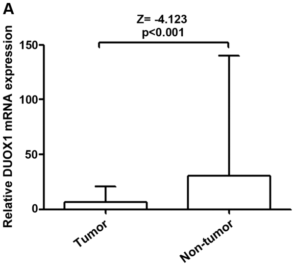
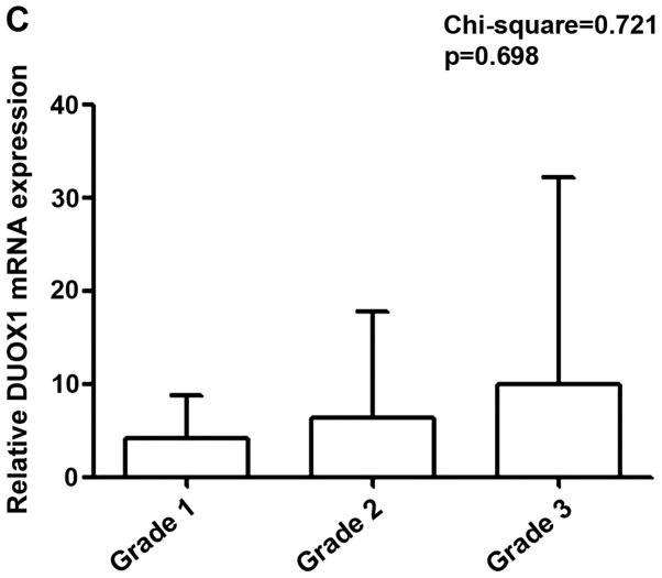
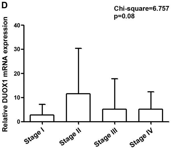
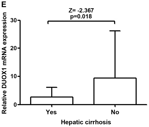
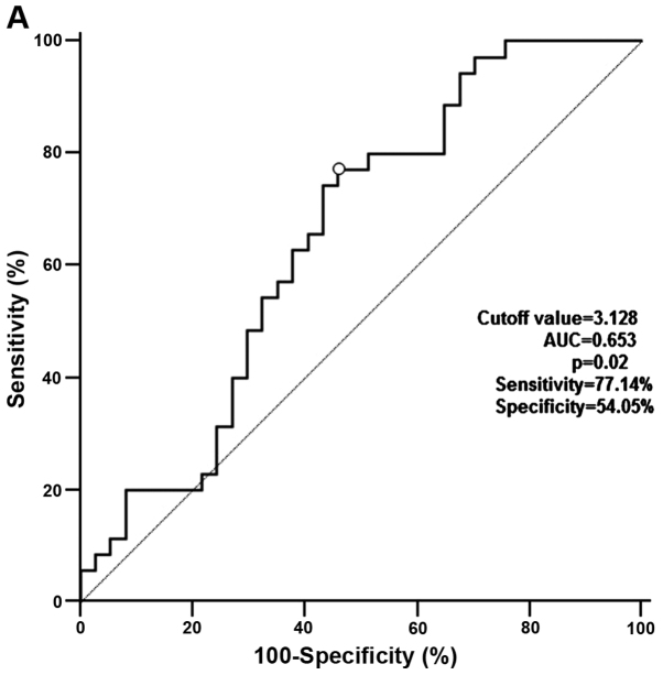
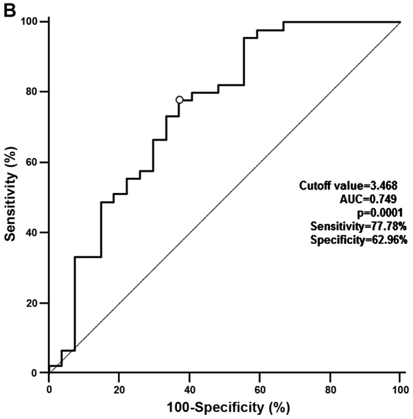
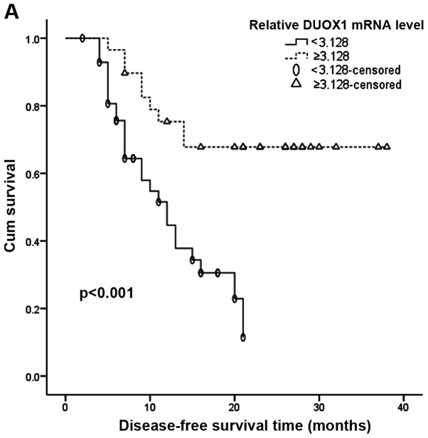
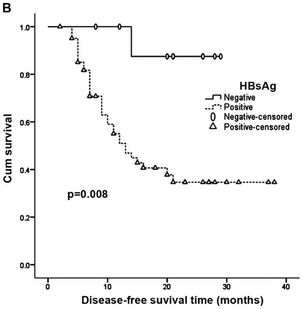
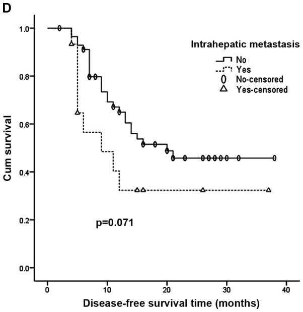
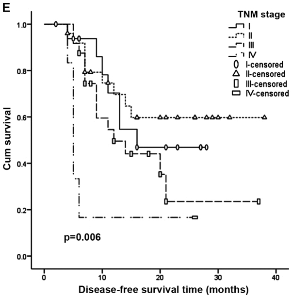
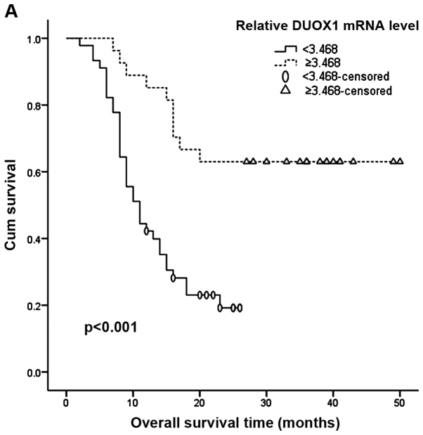
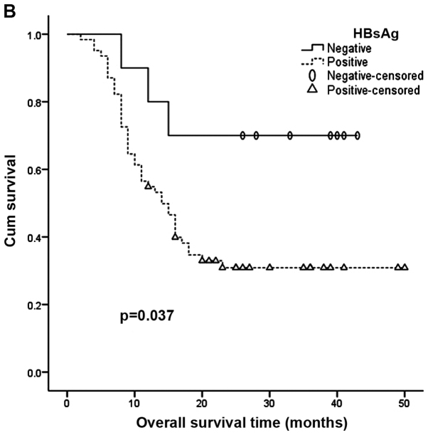
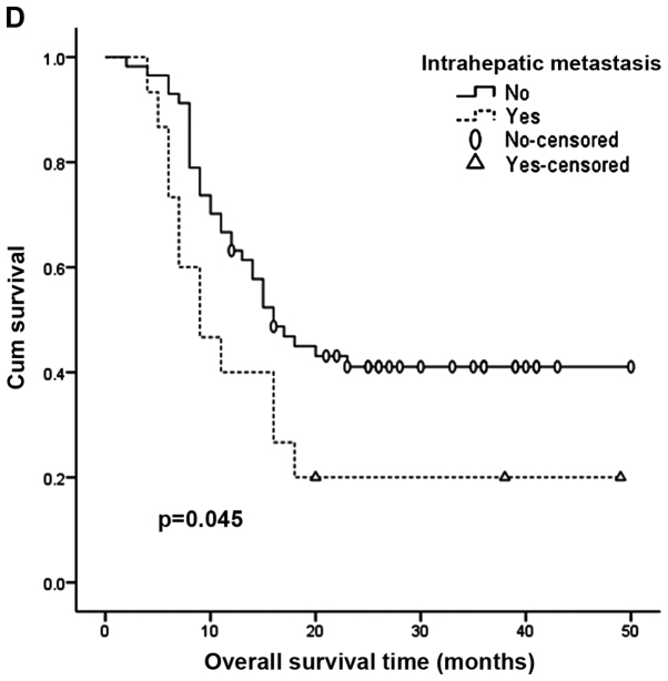
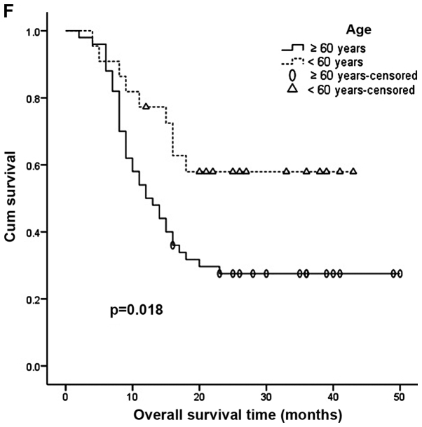
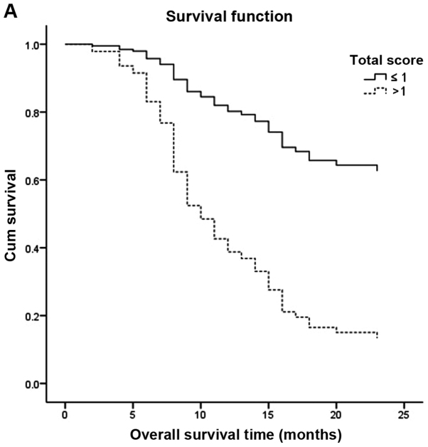
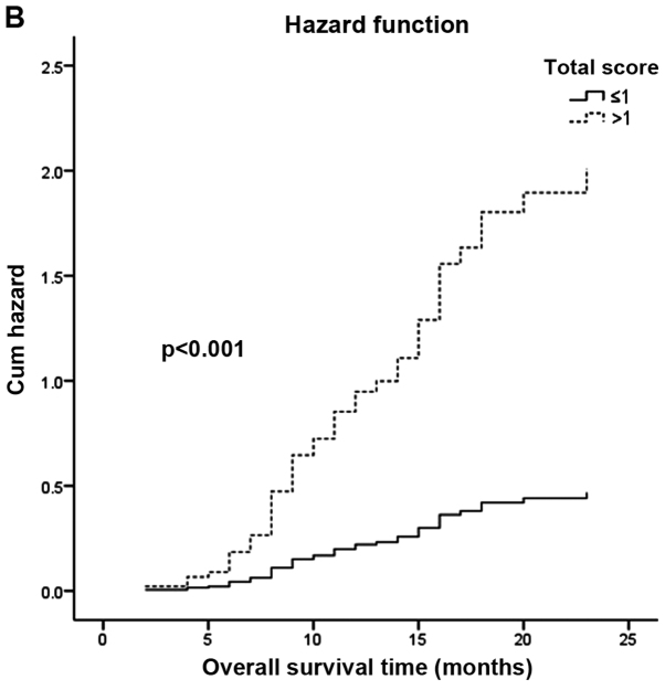
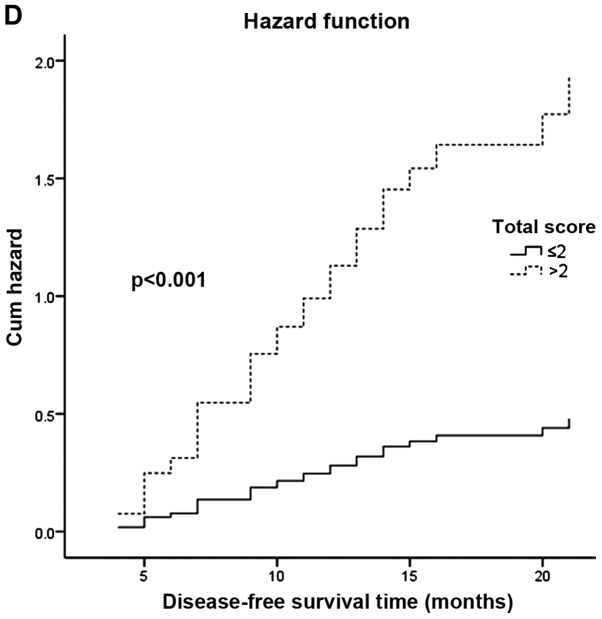
Dual oxidase 1 (DUOX1), which is the main source of reactive oxygen species (ROS) production in the airway, can be silenced in human lung cancer and hepatocellular carcinomas. However, the prognostic value of DUOX1 expression in hepatocellular carcinoma patients is still unclear. We investigated the prognostic value of DUOX1 expression in liver cancer patients. DUOX1 mRNA expression was determined in tumor tissues and non-tumor tissues by real‑time PCR. For evaluation of the prognostic value of DUOX1 expression, Kaplan-Meier method and Cox's proportional hazards model (univariate analysis and multivariate analysis) were employed. A simple risk score was devised by using significant variables obtained from the Cox's regression analysis to further predict the HCC patient prognosis. We observed a reduced DUOX1 mRNA level in the cancer tissues in comparison to the non‑cancer tissues. More importantly, Kaplan-Meier analysis showed that patients with high DUOX1 expression had longer disease-free survival and overall survival compared with those with low expression of DUOX1. Cox's regression analysis indicated that DUOX1 expression, age, and intrahepatic metastasis may be significant prognostic factors for disease-free survival and overall survival. Finally, we found that patients with total scores of >2 and >1 were more likely to relapse and succumb to the disease than patients whose total scores were ≤2 and ≤1. In conclusion, DUOX1 expression in liver tumors is a potential prognostic tool for patients. The risk scoring system is useful for predicting the survival of liver cancer patients after tumor resection.


Figures

Figure 1
Figure 1
Clinicopathologic features and expression of DUOX1. The expression of DUOX1 in HCC tissues and adjacent non-tumor tissues was determined by real-time PCR. (A) Seventy-two pairs of samples were obtained from liver tissues, including tumor tissues and adjacent non-tumor tissues; p-value according to the Mann-Whitney U-test. (B) The expression of DUOX1 mRNA in HBsAg+ and HBsAg groups (p-value according to the Mann-Whitney U-test). (C) The expression of DUOX1 mRNA in different histological grades (three-tier grading scheme) of primary HCC tissues (p-value according to the Kruskal-Wallis test). (D) The expression of DUOX1 mRNA in different TNM stages of primary HCC tissues (p-value according to the Kruskal-Wallis test). (E) The expression of DUOX1 mRNA in hepatic cirrhosis and non-hepatic cirrhosis groups (p-value according to the Mann-Whitney U-test).
Figure 1
Figure 1
Clinicopathologic features and expression of DUOX1. The expression of DUOX1 in HCC tissues and adjacent non-tumor tissues was determined by real-time PCR. (A) Seventy-two pairs of samples were obtained from liver tissues, including tumor tissues and adjacent non-tumor tissues; p-value according to the Mann-Whitney U-test. (B) The expression of DUOX1 mRNA in HBsAg+ and HBsAg groups (p-value according to the Mann-Whitney U-test). (C) The expression of DUOX1 mRNA in different histological grades (three-tier grading scheme) of primary HCC tissues (p-value according to the Kruskal-Wallis test). (D) The expression of DUOX1 mRNA in different TNM stages of primary HCC tissues (p-value according to the Kruskal-Wallis test). (E) The expression of DUOX1 mRNA in hepatic cirrhosis and non-hepatic cirrhosis groups (p-value according to the Mann-Whitney U-test).
Figure 2
Figure 2
ROC curves of DUOX1 expression to indentify the cut-off value of the relative DUOX1 mRNA level. (A) ROC curve of DUOX1 expression for disease-free survival. (B) ROC curve of DUOX1 expression for overall survival.
Figure 2
Figure 2
ROC curves of DUOX1 expression to indentify the cut-off value of the relative DUOX1 mRNA level. (A) ROC curve of DUOX1 expression for disease-free survival. (B) ROC curve of DUOX1 expression for overall survival.
Figure 3
Figure 3
Impact of the clinicopathologic features on patient disease-free survival (DFS) after radical resection for HCC was evaluated using the Kaplan-Meier method (p-value according to the log-rank test). (A) Patients with high DUOX1 expression tended to have longer DFS compared with those with low DUOX1 expression. (B) HBsAg expression, (E) TNM stage and (F) age were significantly correlated with DFS. (C) Histological grade and (D) intrahepatic metastasis were not correlated with DFS.
Figure 3
Figure 3
Impact of the clinicopathologic features on patient disease-free survival (DFS) after radical resection for HCC was evaluated using the Kaplan-Meier method (p-value according to the log-rank test). (A) Patients with high DUOX1 expression tended to have longer DFS compared with those with low DUOX1 expression. (B) HBsAg expression, (E) TNM stage and (F) age were significantly correlated with DFS. (C) Histological grade and (D) intrahepatic metastasis were not correlated with DFS.
Figure 4
Figure 4
Impact of the clinicopathologic features on patient overall survival (OAS) after radical resection for HCC was evaluated using the Kaplan-Meier method (p-value according to the log-rank test). (A) Patients with high DUOX1 expression tended to have longer OAS compared with those with low DUOX1 expression. (B) HBsAg expression, (D) intrahepatic metastasis and (F) age were significantly correlated with OAS. (C) Histological grade and (E) TNM stage were not correlated with OAS.
Figure 4
Figure 4
Impact of the clinicopathologic features on patient overall survival (OAS) after radical resection for HCC was evaluated using the Kaplan-Meier method (p-value according to the log-rank test). (A) Patients with high DUOX1 expression tended to have longer OAS compared with those with low DUOX1 expression. (B) HBsAg expression, (D) intrahepatic metastasis and (F) age were significantly correlated with OAS. (C) Histological grade and (E) TNM stage were not correlated with OAS.
Figure 5
Figure 5
ROC curves with simplified risk score to predict the HCC patient prognosis. (A) ROC curve with simplified risk score to predict the recurrence of HCC in patients after surgery. (B) ROC curve with simplified risk score to predict the death of HCC patients after tumor resection.
Figure 5
Figure 5
ROC curves with simplified risk score to predict the HCC patient prognosis. (A) ROC curve with simplified risk score to predict the recurrence of HCC in patients after surgery. (B) ROC curve with simplified risk score to predict the death of HCC patients after tumor resection.
Figure 6
Figure 6
Iimpact of the total scoring system on disease-free survival and overall survival with Cox's regression analysis (p-value was confirmed with Cox proportional hazards model). Patients whose total score was >2 were more likely to relapse (C and D); patients with a total score >1 were apt to succumb to the disease (A and B) than patients whose scores were ≤2 and ≤1.
Figure 6
Figure 6
Iimpact of the total scoring system on disease-free survival and overall survival with Cox's regression analysis (p-value was confirmed with Cox proportional hazards model). Patients whose total score was >2 were more likely to relapse (C and D); patients with a total score >1 were apt to succumb to the disease (A and B) than patients whose scores were ≤2 and ≤1.

References

    1. Yang JD, Roberts LR. Hepatocellular carcinoma: A global view. Nat Rev Gastroenterol Hepatol. 2010;7:448–458. doi: 10.1038/nrgastro.2010.100. - DOI - PMC - PubMed
    1. Sakamoto M. Early HCC: Diagnosis and molecular markers. J Gastroenterol. 2009;44(Suppl 19):108–111. doi: 10.1007/s00535-008-2245-y. - DOI - PubMed
    1. Roy K, Wu Y, Meitzler JL, Juhasz A, Liu H, Jiang G, Lu J, Antony S, Doroshow JH. NADPH oxidases and cancer. Clin Sci (Lond) 2015;128:863–875. doi: 10.1042/CS20140542. - DOI - PubMed
    1. De Deken X, Wang D, Many MC, Costagliola S, Libert F, Vassart G, Dumont JE, Miot F. Cloning of two human thyroid cDNAs encoding new members of the NADPH oxidase family. J Biol Chem. 2000;275:23227–23233. doi: 10.1074/jbc.M000916200. - DOI - PubMed
    1. Fischer H. Mechanisms and function of DUOX in epithelia of the lung. Antioxid Redox Signal. 2009;11:2453–2465. doi: 10.1089/ars.2009.2558. - DOI - PMC - PubMed

MeSH terms