. 2016 Nov 10;35(45):5882-5892.
doi: 10.1038/onc.2016.119. Epub 2016 Apr 25.

Hypoxia-induced oxidative stress promotes MUC4 degradation via autophagy to enhance pancreatic cancer cells survival

Affiliations

Hypoxia-induced oxidative stress promotes MUC4 degradation via autophagy to enhance pancreatic cancer cells survival

S Joshi et al. Oncogene. .

Abstract

Pancreatic cancer (PC) and associated pre-neoplastic lesions have been reported to be hypoxic, primarily due to hypovascular nature of PC. Though the presence of hypoxia under cancerous condition has been associated with the overexpression of oncogenic proteins (MUC1), multiple emerging reports have also indicated the growth inhibitory effects of hypoxia. In spite of being recognized as the top-most differentially expressed and established oncogenic protein in PC, MUC4 regulation in terms of micro-environmental stress has not been determined. Herein, for the first time, we are reporting that MUC4 protein stability is drastically affected in PC, under hypoxic condition in a hypoxia inducible factor 1α (HIF-1α)-independent manner. Mechanistically, we have demonstrated that hypoxia-mediated induction of reactive oxygen species (ROS) promotes autophagy by inhibiting pAkt/mTORC1 pathway, one of the central regulators of autophagy. Immunohistofluorescence analyses revealed significant negative correlation (P-value=0.017) between 8-hydroxy guanosine (8-OHG) and MUC4 in primary pancreatic tumors (n=25). Moreover, we found pronounced colocalization between MUC4 and LAMP1/LC3 (microtubule-associated protein 1A/1B-light chain 3) in PC tissues and also observed their negative relationship in their expression pattern, suggesting that areas with high autophagy rate had less MUC4 expression. We also found that hypoxia and ROS have negative impact on overall cell growth and viability, which was partially, though significantly (P<0.05), rescued in the presence of MUC4. Altogether, hypoxia-mediated oxidative stress induces autophagy in PC, leading to the MUC4 degradation to enhance survival, possibly by offering required metabolites to stressed cells.


Conflict of interest statement

The authors declare no conflict of interest.

Figures

Figure 1
Figure 1
MUC4 is negatively regulated by hypoxia in PC cell lines. (a) CAPAN1, CD18/HPAF and T3M4 cells were cultured under normoxia or hypoxic (1% O2) conditions for 24 h. Following treatment, lysates were collected and western blots were performed. Protein expression of MUC4 and HIF-1α was analyzed by 2% agarose and 10% polyacrylamide gel-based electrophoresis, respectively. (b) CD18/HPAF cells were grown on coverslips followed by 24 h incubation under normoxia or hypoxia. After the completion of treatment, cells were fixed, permeabilized and then subjected to immunofluorescence experiment to observe changes in the expression of MUC1 and MUC4. (c) Prolong hypoxia treatment was given to CD18/HPAF cells for 72 and 96 h, and the expression of MUC4 and MUC1 was analyzed. (d) qRT-PCR experiment was performed to detect changes in the mRNA expression levels of MUC4 in hypoxia-treated and untreated CD18/HPAF, T3M4 and CAPAN1 PC cell lines (ns stands for no significant difference, scale bar = 20 μM).
Figure 2
Figure 2
HIF-1α-independent mechanisms have a predominant role in hypoxia-mediated suppression of MUC4 expression. (a) After transient knock down of HIF-1α expression, CAPAN1 cells were incubated under 1% hypoxic conditions for 24 h. Following treatment, total protein was isolated and immunoblot was performed to observe the effect of HIF-1α silencing on MUC4 expression under both hypoxic and normoxic conditions. (b) CD18/HPAF and CAPAN1 cells were exposed to different concentrations of YC-1 (10 or 20 μM), an inhibitor of HIF-1α, for 16 h. Immunoblotting was performed to detect changes in MUC4 and HIF-1α expression. (c) CD18/HPAF cells were first pre-treated with MG132 (10 μM) for 30 min. Following pre-treatment, cells were incubated under 1% hypoxic conditions for 4, 6 and 8 h in the presence of MG132. Inhibition of ubiquitin-proteasome pathway, failed to rescue MUC4 degradation, whereas, HIF-1α protein which is known to be degraded solely by proteasome pathway was stabilized upon MG132 treatment under both normoxic and hypoxic conditions. (d) Similar to MG132, CD18/HPAF cells were pre-treated with CHX (50 μg/ml) for 30 min followed by 1% hypoxia treatment for 2, 4 and 6 h. 2% agarose gel electrophoresis was performed to see the effect of these inhibitor treatments on MUC4 expression in the presence or absence of hypoxia. We observed that CHX treatment significantly reduces the levels of MUC4 under hypoxic condition, compared to their respective controls, confirming the negative effect of hypoxia on MUC4 protein stability. (e) Representative images obtained from normal colon and PC tissues (from three different patients) showing MUC4 and HIF-1α co-expression (scale bar = 20 μM).
Figure 3
Figure 3
MUC4 expression is negatively regulated by hypoxia induced ROS. (a) CD18/HPAF cells were treated with NAC in the presence and absence of hypoxia for 24 h. Western blot was performed to analyze alteration in the expression of MUC4 and HIF-1α. (b) Flow cytometry was performed to measure DCFDA fluorescence in order to detect changes in ROS levels in CD18/HPAF cells upon NAC treatment in the presence and absence of hypoxia. (c, d) After 12 h of serum starvation, CD18/HPAF and CAPAN1 cells were treated with α-TS for 24 h at indicated concentrations. Following, MUC4 expression was analyzed by 2% agarose gel electrophoresis. (e, f) CAPAN1 cells and CD18/HPAF cells were treated with H2O2 followed by MUC4 expression analysis. Immunofluorescence experiment was performed to further confirm the effect of hypoxia and exogenous ROS on MUC4 at protein level in the presence and absence of ROS neutralizer, NAC (scale bar = 20 μM).
Figure 4
Figure 4
Hypoxia-mediated ROS production induces autophagy, which leads to reduced MUC4 stability. (a) Cell lysates of CD18/HPAF and CAPAN1 were collected after 24 h incubation with or without 1% hypoxia to analyze the expression of LC3-I and II by immunoblot analysis. (b) CAPAN1 cells were treated with increasing concentrations of H2O2 to observe the effect of oxidative stress on autophagy. (c) To further substantiate that presence of oxidative stress induces autophagy, CD18/HPAF cell line was treated with 40 and 80 μM of H2O2 followed by the analysis of LC3 and p62 levels, using immunoblotting. (d) CAPAN1 cells were treated with 10, 20 and 50 nM of RAP, an autophagy inducer, for 24 h. Cell lysates were prepared to analyze the expression of MUC4 and LC3. (e) CAPAN1 cells were treated with VB (10 μg/ml) for 24 h under hypoxic conditions to observe the effect of autophagy inhibition on MUC4 expression. (f) Additionally, confocal microscopy revealed that inhibition of autophagy due to VB treatment leads to increased expression and retention of MUC4 in LC3-positive vesicles. The bar graph is showing the Pearson correlation coefficient between MUC4 and LC3 colocalization in VB-treated and untreated CD18/HPAF cells. (g) Confocal image demonstrating significant colocalization between MUC4 and LAMP1 in CAPAN1 cell line. (h) To specifically pinpoint the role of autophagy in MUC4 degradation, we used targeted siRNA oligonucleotides to transiently knock down ATG7 in CD18/HPAF PC cells to inhibit autophagy. Consistently, we observed a significant increase in MUC4 expression upon ATG7 silencing (**P < 0.01: statistically significant, scale bar = 20 μM).
Figure 5
Figure 5
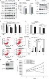
Hypoxia-mediated oxidative stress promotes autophagy by inhibiting pAkt/mTORC1 axis and reduces cell viability. (a) T3M4, CD18/ HPAF and CAPAN1 cells were incubated under 1% hypoxic conditions for 24 h. Following treatment, cell lysates were collected and used for western blotting to observe changes in the proteins expression of HIF-1α, EGFR, pEGFR (Ser1046), Akt, pAkt (Ser473), S6kinase, pS6kinase (Thr389) and p53. (b) Growth kinetics was performed for CD18/HPAF for 24 and 48 h in the presence and absence of 1% hypoxia. (c) To know whether hypoxia-mediated suppression of pAkt and p53 is ROS dependent, CD18/HPAF cells were first pre-treated with NAC (5 mM) for 30 min. Following pre-treatment, cells were incubated under 1% hypoxia. Cell lysates were subsequently collected and immunoblot experiment was performed to analyze Akt, pAkt (Ser473), p53 and MDM2 expression levels. (d) The graphical representation to demonstrate the effect of hypoxia and neutralization of consequently produced ROS (by concomitant treatment with 2.5 mM of NAC) on the proliferation of CD18/HPAF and CAPAN1 cell lines. (e) To explore the role of hypoxia-induced oxidative stress and autophagy on cell death and viability, MTT assay was performed. CD18/HPAF cells were exposed to 1% hypoxia in the presence and absence of NAC (2.5 mM) and CQ (50 μM) for 24 h. Post-treatment, MTT assay was performed and optical density was measured at 570 nm. (f) The graphical representation of Annexin (indicator of early apoptosis) and propidium iodide (PI, indicator of late apoptosis and necrotic cells) staining performed on CD18/HPAF cells treated for 24 h with hypoxia alone, hypoxia followed by NAC (2.5 mM) or CQ (50 μM) treatment for further 12 h. (g) Immunoblot confirming MUC4 knocked down in CAPAN1 cells. (h) The graphical representation to demonstrate the effect of 1, 3 and 5 days of hypoxia treatment on the proliferation of MUC4 kd and scr CAPAN1 cells (LE, low exposure; P<0.05: statistically significant; **P < 0.01: statistically significant; ns, no significant difference).
Figure 6
Figure 6
In vivo validation of MUC4 association with oxidative stress and degradation via lysosomal pathway. (a) Confocal images showing colocalization between MUC4 and lysosomal marker (LAMP1), and thus indicate that MUC4 does enter to lysosomal compartment. In spite of significant colocalization between MUC4 and LAMP1, similar to CAPAN1 cell line, MUC4 and LAMP1 expression pattern was inversely associated under in vivo settings. Tumor cells having higher LAMP1 expression exhibited reduced MUC4 expression in Whipple tissue samples. (b) Representative images of PC tissues stained with MUC4 and oxidative stress marker (or high ROS indicator; 8-OHG). Boxplot showing the significant difference between the MFI observed for 8-OHG in MUC4L (n = 20) and MUCH (n = 16) fields. (c) Representative images obtained from confocal microscopy showing that the presence of oxidative stress does not always correlate with HIF-1α expression, as PC tissue demonstrating high 8-OHG expression had less HIF-1α expression and vice versa. (d) Scatter graph showing the correlation between the MFI levels of HIF-1α and 8-OHG in PC tissue samples (scale bar = 10 μM).
Figure 7
Figure 7
Schematic presentation of the summary of the paper. Hypoxia is induced collaboratively by hypovascularization, desmoplastic reactions and continuous proliferation of tumor cells, which further leads to increased ROS production and generate oxidative stress condition. Produced ROS inhibits the activation of Akt, which further leads to mTORC1 inhibition and induction of autophagy. Induce autophagy facilitates MUC4 degradation. The inhibitors used in this study suppress the activity of different proteins. For example, NAC and α-TOS act as ROS scavenger, RAP inhibits mTORC1 and VB inhibits the fusion of autophagosomes (AP) with lysosomes (L) and thus, prevent the formation of autophagolysosomes that causes MUC4 accumulation.

Similar articles

Cited by

References

    1. Siegel R, Ma J, Zou Z, Jemal A. Cancer statistics, 2014. CA Cancer J Clin. 2014;64:9–29. - PubMed
    1. Sullivan R, Pare GC, Frederiksen LJ, Semenza GL, Graham CH. Hypoxia-induced resistance to anticancer drugs is associated with decreased senescence and requires hypoxia-inducible factor-1 activity. Mol Cancer Ther. 2008;7:1961–1973. - PubMed
    1. Kaur S, Kumar S, Momi N, Sasson AR, Batra SK. Mucins in pancreatic cancer and its microenvironment. Nat Rev Gastroenterol Hepatol. 2013;10:607–620. - PMC - PubMed
    1. Graeber TG, Osmanian C, Jacks T, Housman DE, Koch CJ, Lowe SW, et al. Hypoxia-mediated selection of cells with diminished apoptotic potential in solid tumours. Nature. 1996;379:88–91. - PubMed
    1. Andrianifahanana M, Moniaux N, Schmied BM, Ringel J, Friess H, Hollingsworth MA, et al. Mucin (MUC) gene expression in human pancreatic adenocarcinoma and chronic pancreatitis: a potential role of MUC4 as a tumor marker of diagnostic significance. Clin Cancer Res. 2001;7:4033–4040. - PubMed

MeSH terms

Substances