. 2016 Oct 13;35(41):5388-5399.
doi: 10.1038/onc.2016.76. Epub 2016 Apr 25.

Crosstalk between bone marrow-derived myofibroblasts and gastric cancer cells regulates cancer stemness and promotes tumorigenesis

Affiliations

Crosstalk between bone marrow-derived myofibroblasts and gastric cancer cells regulates cancer stemness and promotes tumorigenesis

Liming Zhu et al. Oncogene. .

Abstract

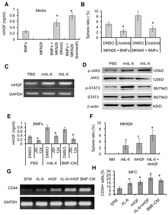
Bone marrow-derived cells have important roles in cancer development and progression. Our previous studies demonstrated that murine bone marrow-derived myofibroblasts (BMFs) enhanced tumor growth. In this study, we investigated the mechanisms of BMF actions. We found that co-injection of BMFs with gastric cancer cells markedly promoted tumorigenesis. Co-cultured BMFs or BMF-conditioned medium (BMF-CM) induced the formation of spheres, which expressed stem cell signatures and exhibited features of self-renewal, epithelial-to-mesenchymal transition and tumor initiation. Furthermore, CD44+ fractions in spheres were able to initiate tumorigenesis and re-establish tumors in serially passaged xenografts. In co-culture systems, BMFs secreted high levels of murine interleukin-6 (IL-6) and hepatocyte growth factor (HGF), whereas cancer cells produced high level of transformation growth factor-β1 (TGF-β1). BMF-CM and IL-6 activated BMFs to produce mHGF, which activated signal transducer and activator of transcription 3 (STAT3) and upregulated TGF-β1 in human cancer cells. In return, cancer cell-CM stimulated BMFs to produce IL-6, which was inhibited by anti-TGF-β1 neutralizing antibody. Blockade of HGF/Met, Janus kinase 2 (JAK2)/STAT3 and TGF-β1 signaling by specific inhibitors inhibited BMF-induced sphere formation. STAT3 knockdown in cancer cells also inhibited BMF-induced sphere formation and tumorigenesis. Moreover, TGF-β1 overexpression in cancer cells was co-related with IL-6 and HGF overexpression in stromal cells in human gastric cancer tissues. Our results show that BMF-derived IL-6/HGF and cancer cell-derived TGF-β1 mediate the interactions between BMFs and gastric cancer cells, which regulate cancer stemness and promote tumorigenesis. Targeting inhibition of the interactions between BMFs and cancer cells may be a new strategy for cancer therapy.


Figures

Fig. 1
Fig. 1. BMFs enhance tumorigenesis in gastric cancer cells
(A) Mouse gastric cancer MFC cells (1 × 10⁴) were injected s.c. alone or together with BMFs (1 × 10⁴) into the both flanks of nude mice. Each group include 5 mice (10 injection sites, n=10). Tumor size was measured weekly. Tumor growth curve was shown accordingly . *P <0.01, compared to MFC cells alone group. (B) The expression of EGFP and α-SMA was determined by immuofluorescence staining in xenograft tumor tissues from mice injected with MFC cells alone or together with BMFs (scale bar, 50 μM). (C-E) Highly tumorigenic gastric cancer MKN45 cells, weakly tumorigenic MKN28 cells and sorted CD44 MKN45 cells (104 cells) were injected (s.c.) alone or co-injected with BMFs (104) into both flank of mice (each group included 5 mice with 10 injection sites) in each experiment. Tumor growth was monitored each week for 3 months. The rate of tumor formation presented was from 5 mice with 10 injection sites each group (n=10). *P <0.01, compared to MKN45 cells alone group. (F) MKN45 cells were cultured alone or co-cultured with BMFs in SCM in attachment 6-well plates for 2 weeks. (G) Mouse gastric cancer cells MFC were cultured in SCM, BMF-CnM or Co-culture-CM in an ultralow attachment 96-well plate for 2 weeks. Representative sphere photos were taken on day 14 (scale bar, 100 μM). (H) MKN28 (EGFP) cells were cultured alone or with BMFs (EGFP+) for 2 weeks. All representative sphere photos were taken on day 14 (scale bar, 100 μM).
Fig. 2
Fig. 2
BMF-induced spheres exhibit CSC properties. (A) Single spheroid cells isolated from BMF-CM-induced first generation spheres (second panel) were cultured in an ultralow attachment 96-well plate for 2 weeks and formed second generation spheres (third panel). The second generation spheroid cells were cultured in attachment plates and exhibited fibroblast-like morphology (fourth panel) (scale bar, 50 μM). (B) MKN28 cells and single MKN28-CLC-LCs were cultured in a 96-well plate in SCM for 2 weeks. Sphere ratio is means ± SD of 3 independent experiments. *P < 0.01, compared to MKN28 parental cells. (C) Protein expressions of EMT-related genes in indicated cells were determined by Western Blot. (D) MKN28-CSC-LCs and MKN28-parental cells at indicated cell numbers were injected into the flanks of athymic nude mice (n=5). Tumor formation was monitored for 3 months. (E) Sorted CD44+ and CD44 MKN45 cells from MKN45-CSC-LCs at indicated numbers were injected s.c. into the both flanks of nude mice (n=5). Tumor formation was monitored for 3 months. The percentage of mice with tumors was shown. *P < 0.01, compared to CD44 cells. (F) Sorted CD44+ and CD44 tumor cells from xenograft tumors at indicated numbers were injected s.c. into the flanks of nude mice (n=5). Tumor formation was monitored for 3 months. The percentage of mice with tumors is shown.*P < 0.01, compared to CD44 cells. (G) First and second transplants tumor tissues were suffered H&E and CD44 staining.
Fig. 3
Fig. 3
BMF-derived IL-6 induces sphere formation of mouse cancer cells. (A) BMFs were cultured alone or co-cultured with MKN28 cells in serum-free media for 48 hours. The levels of various factors in culture media were measured by antibody array assay and were normalized to positive control, which is the reagents in the kit which is from Ray Biotech Company. Data are expressed as fold inductions from duplicate experiments. (B-C) BMFs were cultured alone, mixed co-cultured, or co-cultured in a transwell system with MKN28 cells (B) and MFC cells (C) for 48 hours. The mIL-6 level in the media was measured by anti-mouse IL-6 ELISA kit. (D) Mouse MFC cells were cultured for 2 weeks with SCM, BMF-CM or Co-Culture-CM that has been pre-incubated with anti-mouse IL-6 neutralizing antibody or control IgG. (E) MFC cells and MKN28 cells were co-cultured with BMFs in the presence or absence of JSI-124 (0.25 μM) for 2 weeks. All sphere ratios are means ± SD of 3 independent experiments. *P < 0.05, compared to the control groups.
Fig. 4
Fig. 4
BMF-derived HGF contributes to the sphere formation of human cancer cells. (A) BMFs were mixed co-cultured or in a transwell system with MKN28 cells in 2 ml of media for 48 hours. The mHGF level in the media was determined by ELISA. (B) MKN28 and MKN45 cells were co-cultured with BMFs in SCM in the presence or absence of crizotinib (1.00 μM) for 2 weeks. *P <0.05, compared to DMSO group. (C) BMFs were treated with rIL-6 for 48 hours and the HGF mRNA expression was determined by RT-PCR. (D) BMFs were treated with rIL-6 for 10 min. The protein expression was determined by Western Blot. (E) BMFs were treated with rhIL-6 or BMF-CM in the presence or absence of JSI-124 (0.25 μM) for 48 hours. mIL-6 level was measured by ELISA. *P < 0.01, compared to the vehicle control. MKN28 cells were treated with rhIL-6 (20 ng/mL), rhHGF (20 ng/mL) or their combination (20 ng/mL, each) in normal media(NM) for 2 weeks. Sphere ratio is means ± SD from 3 independent experiments. *P < 0.05, compared to rhIL-6 and rhHGF treatment alone; #P <0.05, compared to NM group. (G-H) MFC cells were treated with indicated factors or medium for 24 hours. mRNA expression of CD44 was determined by RT-PCR (H) and percentages of CD44+ was determined by FACS. *P < 0.05, compared to Serum-Free Media (SFM) treatment.
Fig. 5
Fig. 5
The activation of STAT3 and Met contributes to BMF-induced sphere formation and tumorigenesis. Human gastric cancer MKN28 cells were cultured in SCM or BMF-CM (A) or rmHGF (20 ng/mL) (B) in indicated time. (C) Cancer cells were treated with BMF-CM in presence or absence of crizotinib (20 μM) for 2 hours. All protein expression was determined by Western Blot. (D) Stable MKN28-STAT3-shRNA and MKN28-Ctrl-shRNA cells were treated with rhIL-6 (20 ng/mL) for 48 hours. Protein expression was detected by Western Blot. (E) Stable MKN28-STAT3-shRNA and MKN28-Ctrl-ShRNA cells were cultured in BMFCM in 96-well plates for 2 weeks. The sphere ratio is the means ± SD of 3 independent experiments. (F) The MKN28-STAT3-shRNA and MKN28-Ctrl-ShRNA cells were injected s.c. alone or together with BMFs into the both flanks of nude mice (n=4 mice, 8 injection sites). The representative photos were taken in week 5 from one of two experiments.
Fig. 6
Fig. 6
MKN28 cells (A), MKN45 (B) or MFC (C) were cultured alone or co-cultured mixedly or in a transwell system with BMFs cells for 48 hours. The hTGF-β1 level in the media was measured by anti-human TGF-β1 ELISA kit. *P < 0.01, compared to cancer cell culture alones. Cancer cell-derived TGF-β1 activates BMFs. (D) The BMFs were treated with rhTGF-β1, MKN28-CM or Co-culture-CM in the presence or absence of anti-human TGF-β1 neutralizing antibody for 48 hours. *P < 0.01, compared to control IgG. #P < .01, compared to NM. (E) BMFs were cultured alone or with MKN28 cells in the presence or absence of SB-505124 for 48 hours. The mIL-6 level was measured by ELISA. *P < 0.01, compared to control groups. (F) MKN28 or MKN45 were co-cultured with BMFs with and without SB-505124 in SCM for 2 weeks. Sphere ratio is the means ± SD of 3 independent experiments. *P < 0.01, compared to control groups.
Fig. 7
Fig. 7
The correlations among expressions of hTGF-β1, hIL-6 and hHGF in human gastric cancer tissues. (A) Expressions of hTGF-β1, HGF and hIL-6 in human gastric cancer tissues array samples were determined by IHC with anti-human TGF-β1, HGF and IL-6 antibodies using. (B-D) Association analysis of mRNA expression of hTGF-β1, hHGF and hIL-6 in NIH TCGA gastric cancer database. (E) A model for the interactions between BMFs and cancer cells through an IL-6/HGF and TGF-β1 signaling loop.

Similar articles

Cited by

References

    1. Double blind controlled phase III multicenter clinical trial with interferon gamma in rheumatoid arthritis. German Lymphokine Study Group. Rheumatol Int. 1992;12:175–185. - PubMed
    1. Beppu H, Mwizerwa ON, Beppu Y, Dattwyler MP, Lauwers GY, Bloch KD, et al. Stromal inactivation of BMPRII leads to colorectal epithelial overgrowth and polyp formation. Oncogene. 2008;27:1063–1070. - PubMed
    1. Bhowmick NA, Neilson EG, Moses HL. Stromal fibroblasts in cancer initiation and progression. Nature. 2004;432:332–337. - PMC - PubMed
    1. Birkenkamp-Demtroder K, Maghnouj A, Mansilla F, Thorsen K, Andersen C, Ãs̃ter B, et al. Repression of KIAA1199 attenuates Wnt-signalling and decreases the proliferation of colon cancer cells. British journal of cancer. 105:552–561. - PMC - PubMed
    1. Calon A, Espinet E, Palomo-Ponce S, Tauriello DV, Iglesias M, Cespedes MV, et al. Dependency of colorectal cancer on a TGF-beta-driven program in stromal cells for metastasis initiation. Cancer Cell. 2012;22:571–584. - PMC - PubMed

Publication types

MeSH terms