

Blastomere removal from cleavage-stage mouse embryos alters placental function, which is associated with placental oxidative stress and inflammation
- PMID: 27109212
- PMCID: PMC4842963
- DOI: 10.1038/srep25023
Blastomere removal from cleavage-stage mouse embryos alters placental function, which is associated with placental oxidative stress and inflammation
Abstract
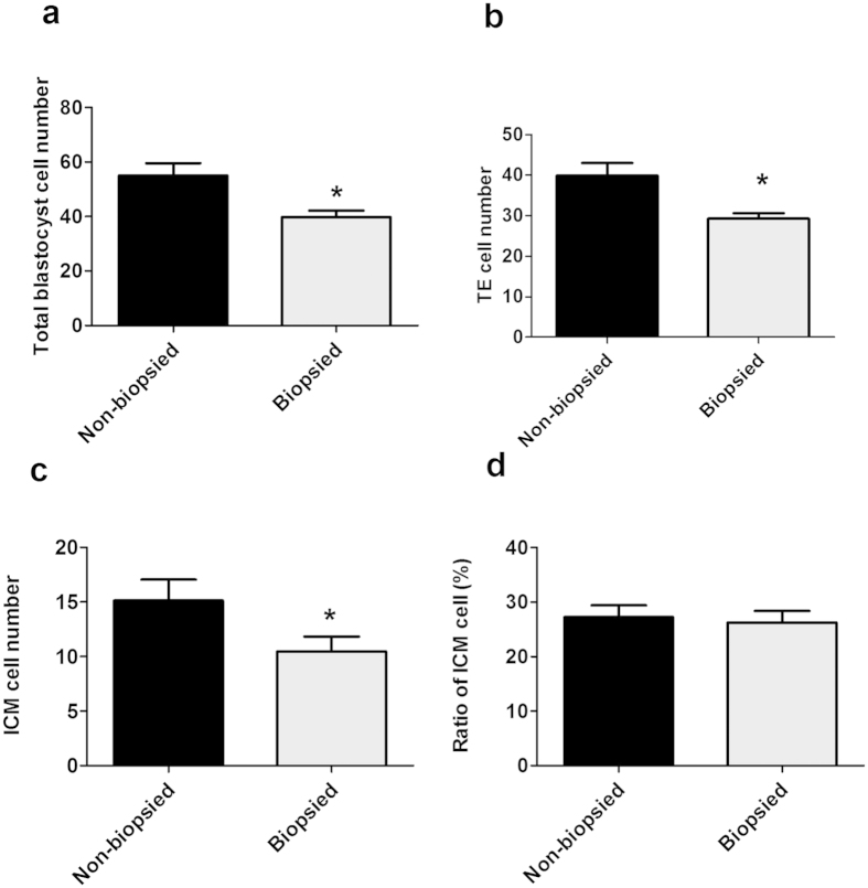
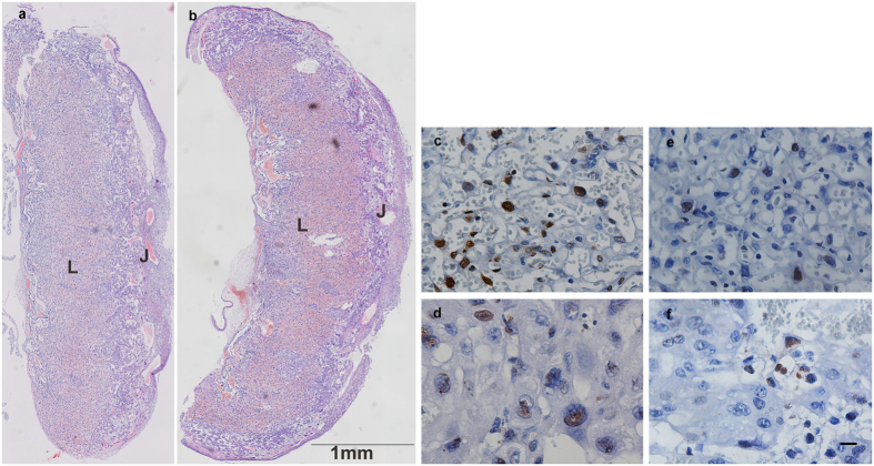
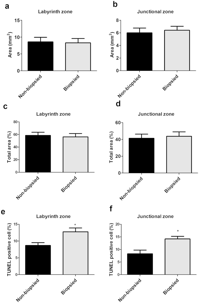
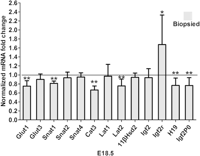
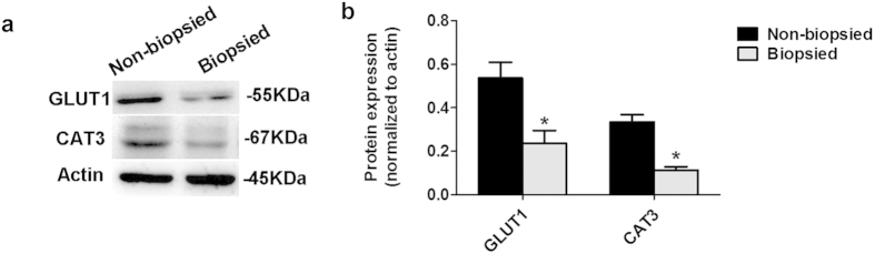
Blastomere biopsy is an essential technique in preimplantation genetic diagnosis (PGD), a screening test that can detect genetic abnormalities of embryos before their transfer into uterus. Our results showed that the weights of fetuses derived from biopsied embryos were lower than that of non-biopsied counterparts at E12.5, E15.5, and E18.5. The ratio of fetal/placental (F/P) weights in the biopsied group was significantly lower than that in the non-biopsied group at E18.5. At E18.5, the mRNAs for selected glucose transporters, system A amino acid transporters, system L amino acid transporters, and imprinted genes were downregulated in the placentae of biopsied group, and the GLUT1 and CAT3 protein levels were decreased too. More apoptotic cells were detected by TUNEL in the placentae of biopsied group. Placentae from biopsied embryos exhibited lower levels of SOD and GSH. Furthermore, the concentration of MDA increased in the placentae from biopsied group. The levels of IL1B, IL6, and TNFA also significantly increased in the placentae of biopsied group. This study suggested that placental function may be sensitive to blastomere biopsy procedures, and placental oxidative stress and inflammation associated with blastomere biopsy may be critical factors of abnormal placental function and further influence the fetal development.
Figures
Similar articles
-
Single blastomere removal from murine embryos is associated with activation of matrix metalloproteinases and Janus kinase/signal transducers and activators of transcription pathways of placental inflammation.Mol Hum Reprod. 2014 Dec;20(12):1247-57. doi: 10.1093/molehr/gau072. Epub 2014 Sep 1. Mol Hum Reprod. 2014. PMID: 25180268 Free PMC article.
-
Blastomere removal from cleavage-stage mouse embryos alters steroid metabolism during pregnancy.Biol Reprod. 2012 Jul 5;87(1):4, 1-9. doi: 10.1095/biolreprod.111.097444. Print 2012 Jul. Biol Reprod. 2012. PMID: 22517623 Free PMC article.
-
Aberrant epigenetic modification in murine brain tissues of offspring from preimplantation genetic diagnosis blastomere biopsies.Biol Reprod. 2013 Nov 21;89(5):117. doi: 10.1095/biolreprod.113.109926. Print 2013 Nov. Biol Reprod. 2013. PMID: 24089199
-
Human embryonic development after blastomere removal: a time-lapse analysis.Hum Reprod. 2012 Jan;27(1):97-105. doi: 10.1093/humrep/der382. Epub 2011 Nov 10. Hum Reprod. 2012. PMID: 22081251
-
Survival and developmental potential of stored human early cleavage stage embryos.Eur J Obstet Gynecol Reprod Biol. 2004 Jul 1;115 Suppl 1:S8-11. doi: 10.1016/j.ejogrb.2004.01.009. Eur J Obstet Gynecol Reprod Biol. 2004. PMID: 15196708 Review.
Cited by
-
Pregnancies through oocyte donation. A mini review of pathways involved in placental dysfunction.Front Med (Lausanne). 2024 Jan 17;11:1338516. doi: 10.3389/fmed.2024.1338516. eCollection 2024. Front Med (Lausanne). 2024. PMID: 38298815 Free PMC article. Review.
-
Sex and Exposure to Postnatal Chlorpyrifos Influence the Epigenetics of Feeding-Related Genes in a Transgenic APOE Mouse Model: Long-Term Implications on Body Weight after a High-Fat Diet.Int J Environ Res Public Health. 2020 Dec 29;18(1):184. doi: 10.3390/ijerph18010184. Int J Environ Res Public Health. 2020. PMID: 33383760 Free PMC article.
-
A transcriptomic analysis in mice following a single dose of ibogaine identifies new potential therapeutic targets.Transl Psychiatry. 2024 Jan 19;14(1):41. doi: 10.1038/s41398-024-02773-7. Transl Psychiatry. 2024. PMID: 38242896 Free PMC article.
-
A Non-invasive Chromosome Screening Strategy for Prioritizing in vitro Fertilization Embryos for Implantation.Front Cell Dev Biol. 2021 Aug 9;9:708322. doi: 10.3389/fcell.2021.708322. eCollection 2021. Front Cell Dev Biol. 2021. PMID: 34434931 Free PMC article.
-
Trophectoderm biopsy is associated with adverse obstetric outcomes rather than neonatal outcomes.BMC Pregnancy Childbirth. 2023 Mar 4;23(1):141. doi: 10.1186/s12884-023-05466-z. BMC Pregnancy Childbirth. 2023. PMID: 36870973 Free PMC article.
References
-
- Steptoe P. C. & Edwards R. G. Birth after the reimplantation of a human embryo. Lancet 2, 366 (1978). - PubMed
-
- Servick K. Unsettled questions trail IVF’s success. Science 345, 744–746 (2014). - PubMed
-
- McMillen I. C. & Robinson J. S. Developmental origins of the metabolic syndrome: prediction, plasticity, and programming. Physiol Rev 85, 571–633 (2005). - PubMed
Publication types
MeSH terms
LinkOut - more resources
Full Text Sources
Other Literature Sources
Medical
Miscellaneous
