. 2016 Aug;136(8):1692-1700.
doi: 10.1016/j.jid.2016.03.041. Epub 2016 Apr 22.

A Powerful Mitochondria-Targeted Iron Chelator Affords High Photoprotection against Solar Ultraviolet A Radiation

Affiliations

A Powerful Mitochondria-Targeted Iron Chelator Affords High Photoprotection against Solar Ultraviolet A Radiation

Olivier Reelfs et al. J Invest Dermatol. 2016 Aug.

Abstract

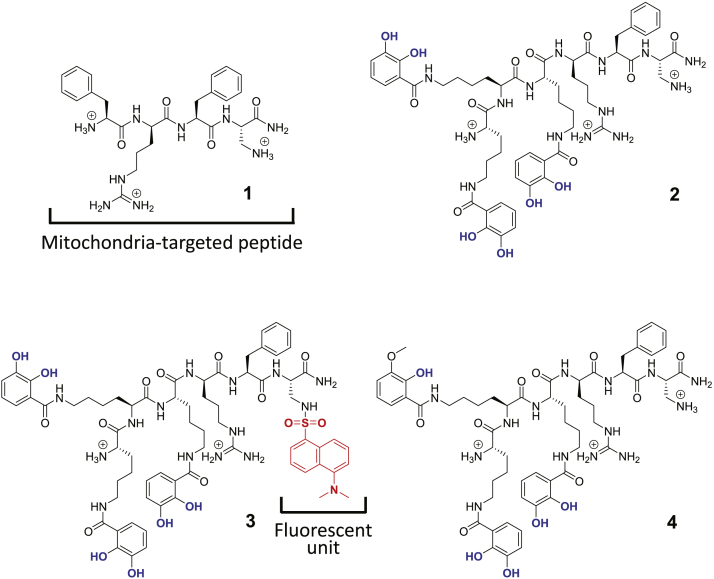
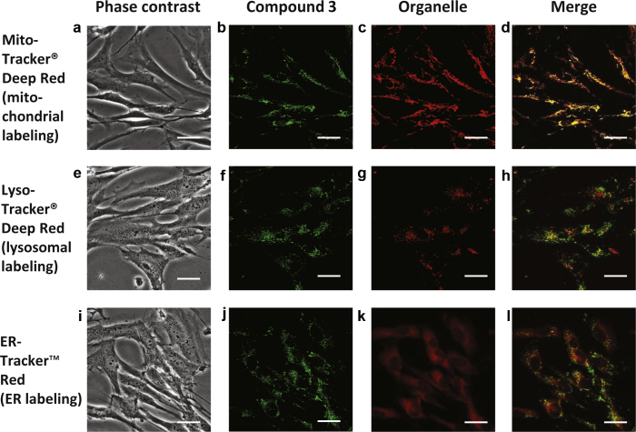
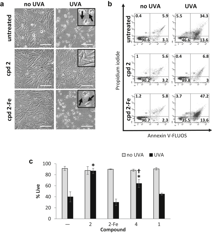
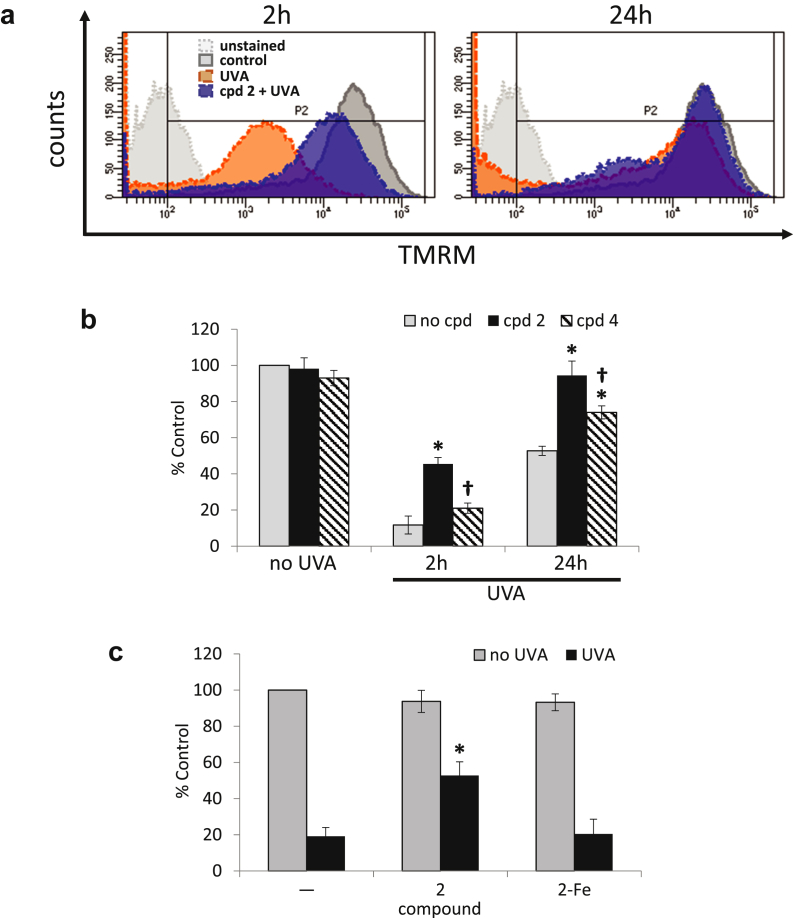
Mitochondria are the principal destination for labile iron, making these organelles particularly susceptible to oxidative damage on exposure to ultraviolet A (UVA, 320-400 nm), the oxidizing component of sunlight. The labile iron-mediated oxidative damage caused by UVA to mitochondria leads to necrotic cell death via adenosine triphosphate depletion. Therefore, targeted removal of mitochondrial labile iron via highly specific tools from these organelles may be an effective approach to protect the skin cells against the harmful effects of UVA. In this work, we designed a mitochondria-targeted hexadentate (tricatechol-based) iron chelator linked to mitochondria-homing SS-like peptides. The photoprotective potential of this compound against UVA-induced oxidative damage and cell death was evaluated in cultured primary skin fibroblasts. Our results show that this compound provides unprecedented protection against UVA-induced mitochondrial damage, adenosine triphosphate depletion, and the ensuing necrotic cell death in skin fibroblasts, and this effect is fully related to its potent iron-chelating property in the organelle. This mitochondria-targeted iron chelator has therefore promising potential for skin photoprotection against the deleterious effects of the UVA component of sunlight.


Figures

Figure 1
Figure 1
Structures of compounds 1–4. The structures of the compounds investigated are depicted with their ionized functions at pH 7.4. Iron-binding functions are indicated in blue. The dansyl (Dns) fluorophore is indicated in red.
Figure 2
Figure 2
Microscopy images of subcellular localization studies of the DNS-labeled peptide-chelator H-Hbl-Hbl-Hbl-r-F-DAP(Dns)-NH2 (compound 3). Representative microscopy images are shown, of cells stained with compound 3 in combination with markers for mitochondrial (a–d), lysosomal (e–h), and ER (i–l) compartments. Fluorescence data were collected and analyzed as described in the Materials and Methods section. Green (b, f, j) and red (c, g, k) fluorescence data were merged together (d, h, l) to identify colocalization in yellow. The phase contrast images (a, e, i) corresponding to the fluorescence data are also shown. Scale bar = 10 μm. Dns, dansyl; ER, endoplasmic reticulum.
Figure 3
Figure 3
Compound 2 protects FEK4 cells from UVA-induced cell death. Cells were treated with compound 2 (cpd 2) alone or as a complex with iron (cpd 2-Fe) or UVA or combinations thereof. (a) Bright-field images were captured 24 hours after treatment. Swelling (arrows in inset) is indicative of cell death by necrosis and is visible after UVA treatment alone or in combination with cpd 2-Fe. Scale bar = 50 μm. (b) Cells treated as described above were processed for flow cytometry analysis as described in the Materials and Methods section. Dot plots of a representative experiment are shown. Live cells are defined as Annexin V-negative/PI-negative (lower left-hand quadrant). (c) Bar chart of flow cytometry results, expressed as means ± SD of percentage live cells from 3 to 5 experiments. *Significantly different (P < 0.05) from UVA alone. Significantly different (P < 0.05) from cpd 2+UVA. UVA, ultraviolet A.
Figure 4
Figure 4
Compound 2 significantly reduces UVA-induced damage to mitochondria membrane. (a) Representative fluorescence intensity profiles of samples stained with TMRM at 2 and 24 hours after UVA irradiation as described in the Materials and Methods section. (b) Bar chart of the results of TMRM staining experiments. Fluorescence median intensities (MFIs) collected over the region delineated in panel a as P2 are expressed as percentage of untreated control. *Significantly different (P < 0.05) from the corresponding UVA-treated sample. Significantly different (P < 0.05) from the corresponding cpd 2+UVA-treated sample. (c) FEK4 cells were pretreated with either compound 2 alone or as a complex with iron, before UVA irradiation and ATP production was measured as described in the Materials and Methods section. Results are expressed as percentage of untreated control. *Significantly different (P < 0.05) from the UVA alone sample. ATP, adenosine triphosphate; TMRM, tetramethylrhodamine methyl ester; UVA, ultraviolet A.

References

    1. Abbate V., Reelfs O., Hider R.C., Pourzand C. Design of novel fluorescent mitochondria targeted peptides with iron-selective sensing activity. Biochem J. 2015;469:357–366. - PubMed
    1. Abbate V., Reelfs O., Kong X., Pourzand C., Hider R.C. Dual selective iron chelating probes with a potential to monitor mitochondrial labile iron pools. Chem Commun (Camb) 2015;2:784–787. - PubMed
    1. Al-Qenaei A., Yiakouvaki A., Reelfs O., Santambrogio P., Levi S., Hall N.D. Role of intracellular labile iron, ferritin, and antioxidant defence in resistance of chronically adapted Jurkat T cells to hydrogen peroxide. Free Radic Biol Med. 2014;68:87–100. - PMC - PubMed
    1. Aroun A., Zhong J.L., Tyrrell R.M., Pourzand C. Iron, oxidative stress and the example of solar ultraviolet A radiation. Photochem Photobiol Sci. 2012;11:118–134. - PubMed
    1. Basu-Modak S., Ali D., Gordon M., Polte T., Yiakouvaki A., Pourzand C. Suppression of UVA-mediated release of labile iron by epicathecin-A link to lysosomal protection. Free Radic Biol Med. 2006;41:1197–1204. - PubMed

Publication types

MeSH terms