Stereoscopic Display of the Peripheral Nerves at the Elbow Region Based on MR Diffusion Tensor Imaging with Multiple Post-Processing Methods
- PMID: 27110337
- PMCID: PMC4836049
- DOI: 10.5812/iranjradiol.22144
Stereoscopic Display of the Peripheral Nerves at the Elbow Region Based on MR Diffusion Tensor Imaging with Multiple Post-Processing Methods
Abstract
Background: Peripheral nerves at the elbow region are prone to entrapment neuropathies and injuries. To make accurate assessment, clinicians need stereoscopic display of the nerves to observe them at all angles.
Objectives: To obtain a stereoscopic display of the peripheral nerves at the elbow region based on magnetic resonance (MR) diffusion tensor imaging (DTI) data using three post-processing methods of volume rendering (VR), maximum intensity projection (MIP), and fiber tractography, and to evaluate the difference and correlation between them.
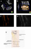
Subjects and methods: Twenty-four elbows of 12 healthy young volunteers were assessed by 20 encoding diffusion direction MR DTI scans. Images belonging to a single direction (anterior-posterior direction, perpendicular to the nerve) were subjected to VR and MIP reconstruction. All raw DTI data were transferred to the Siemens MR workstation for fiber tractography post-processing. Imaging qualities of fiber tractography and VR/MIP were evaluated by two observers independently based on a custom evaluation scale.
Results: Stereoscopic displays of the nerves were obtained in all 24 elbows by VR, MIP, and fiber tractography post-processing methods. The VR/MIP post-processing methods were easier to perform compared to fiber tractography. There was no significant difference among the scores of fiber tracking and VR/MIP reconstruction for single direction. The imaging quality scores of fiber tractography and VR/MIP were significantly correlated based on intraclass correlation coefficient (ICC) analysis (ICC ranged 0.709 - 0.901), which suggested that the scores based on fiber tractography and VR/MIP for the same sample were consistent. Inter- and intraobserver agreements were good to excellent.
Conclusion: Stereoscopic displays of the peripheral nerves at the elbow region can be achieved by using VR, MIP, and fiber tracking post-processing methods based on raw DTI images. VR and MIP reconstruction could be used as preview tools before fiber tracking to determine whether the raw images are satisfactory.
Keywords: Diffusion Tensor Imaging; Magnetic Resonance Imaging; Peripheral Nerves.
Figures


References
LinkOut - more resources
Full Text Sources
Other Literature Sources
